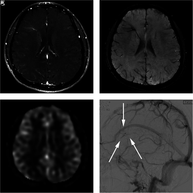
FIG 3.
A 29-year-old man presenting with an AVM in the left occipital lobe (case 11). A, Contrast-enhanced T1-weighted axial MR imaging shows a DVA-like lesion in the right frontal lobe. B, SWI shows hypointense signal in the lesion. C, An ASL quantitative CBF image demonstrates no identifiable signal corresponding to the lesion. D, On DSA, the lesion is first visualized in the late venous phase (arrows), as is typically seen in a classic DVA.

