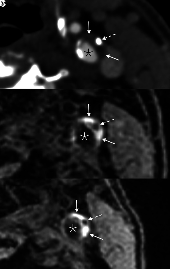
FIG 2.

Example of persistent IPH in a 71-year-old man who presented with acute disorientation and unsteady gait. MR imaging of the brain at the time of admission (not shown) demonstrated multiple acute left cerebral infarcts. Axial CTA image (A) shows a mixed calcified (dashed arrow) and soft (solid arrows) plaque in the left ICA. Corresponding MPRAGE image (B) demonstrates IPH throughout the soft plaque components (solid arrows); the focal calcification is also noted (dashed arrow). The patient was started on dual antiplatelet therapy (aspirin and clopidogrel). One year later (C), the appearance of the IPH (solid arrows) and calcification (dashed arrow) was unchanged. Asterisks denote the vessel lumen.
