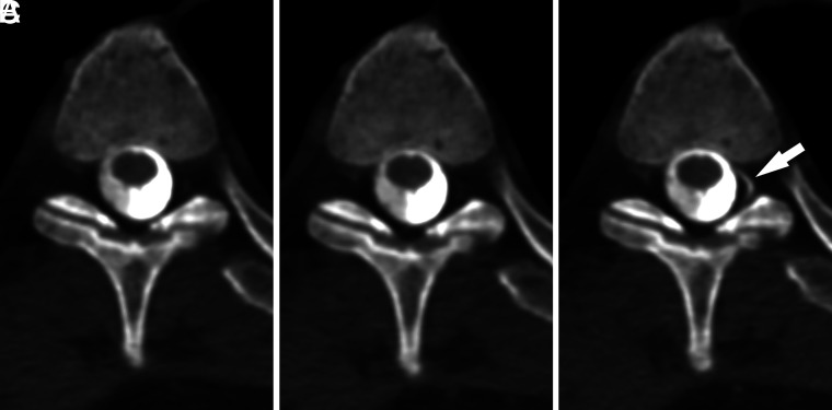
FIG 3.
A left T3 CVF best visualized during the Valsalva maneuver. Axial MIP images from CTM performed on a patient with SIH are shown during maximum suspended inspiration (A), resisted inspiration (B), and the Valsalva maneuver (C). The CVF originating at the T3 level on the left (arrow) is best visualized during the Valsalva maneuver. The presence of a CVF was suspected on the basis of faint hyperdensity in the same location on the initial maximum suspended inspiration image.

