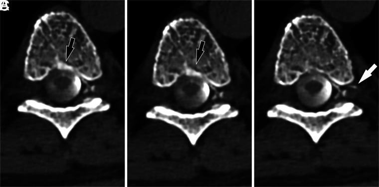
FIG 4.
A left T7 CVF drainage pattern shifts with varying respiratory phases. Axial images from CTM performed on a patient with SIH are shown during maximum suspended inspiration (A), resisted inspiration (B), and the Valsalva maneuver (C). The primary drainage of the CVF is into the internal epidural venous plexus, but during inspiratory phases, a component of drainage is also seen into the basivertebral venous plexus (black arrows). This drainage decreases during the Valsalva maneuver, with new drainage visible into the external epidural venous plexus via an intervertebral vein (white arrow).

