Figure 2.
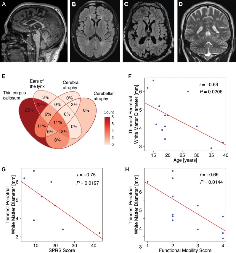
Analysis of the neuroimaging spectrum of HSP-ZFYVE26 (SPG15) delineates several core features. Key neuroimaging findings in HSP-ZFYVE26 include thinning of the corpus callosum which predominantly affects the anterior parts (A, sagittal T1-weighted image of an 18-year-old patient). (B and C) White matter signal changes include a classic ‘ears of the lynx’ sign as well as diffuse periventricular white matter signal changes (B, axial T2-FLAIR image of an 18-year-old patient). Cerebral volume loss and enlarged lateral ventricles are seen in a subset of patients (C, axial T2-FLAIR image of a 22-year-old patient). (D) Cerebellar volume loss is uncommon and usually presents with mild prominence of the cerebellar fissures (coronal T2-weighted image of an 18-year-old patient). (E) Venn diagram of key MRI findings in HSP-ZFYVE26. This consists of (i) thinning of the corpus callosum; (ii) abnormal signal of the forceps minor consistent with an ‘ears of the lynx’ appearance; (iii) cerebral volume loss; and (iv) cerebellar volume loss. (F–H) Correlation analysis of MRI findings and clinical characteristics or motor function scores. Red lines represent linear regression lines. Periventricular white matter, approximated using the thinnest periatrial white matter diameter, inversely correlates with age, the SPRS and the Four Stage Functional Mobility Score as an indicator of motor impairment and associated complications.

