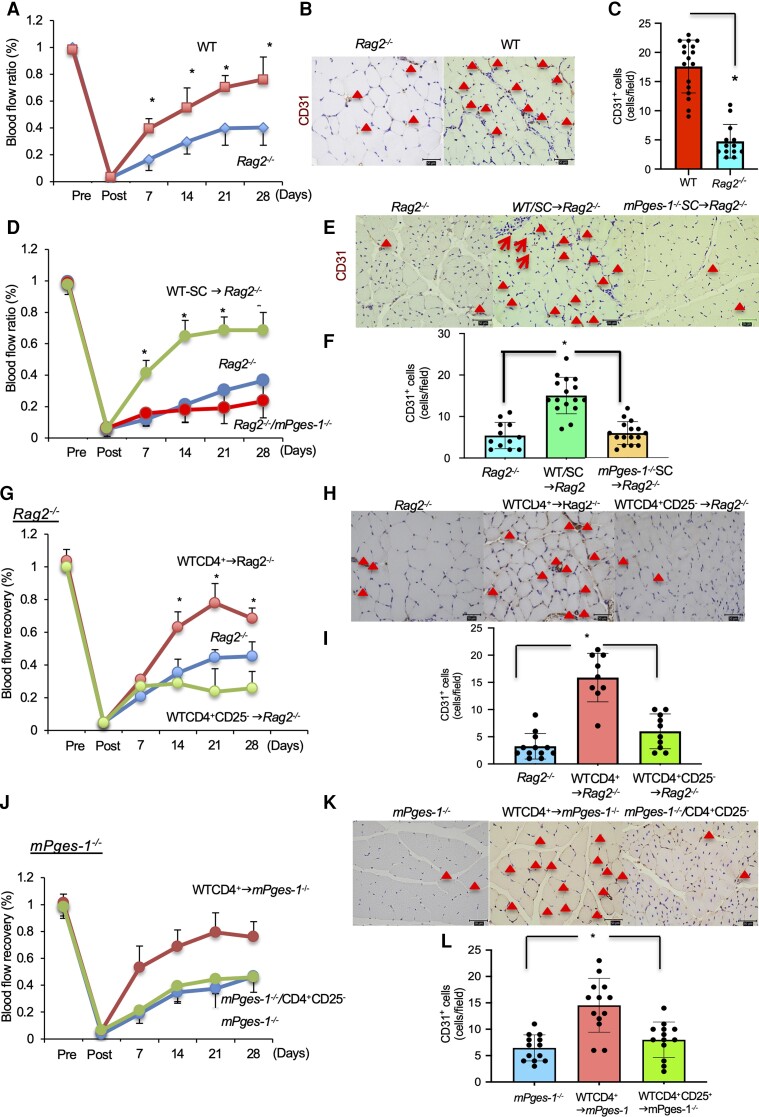
Figure 3.
Transplantation of Tregs induced recovery from ischaemia via mPGES-1. (A) Transplantation of WT-derived CD4+ cells into Rag2−/− recipient mice enhanced recovery from ischaemia. (B) CD31 staining of the ischaemic limb 28 days after ligation. Triangles indicate CD31+ cells. Scale bar, 50 μm. (C) Number of CD31+ cells in ischaemic muscle tissue 28 days after ligation. P < 0.05 vs. PBS, Student’s t-test. (D) Rag2−/− recipient mice transplanted with mPges-1−/−-derived CD4+ cells delayed blood flow recovery. Data are means ± SD of the n = 4 mice/group. *P < 0.05 vs. WT-SC→Rag2−/−, repeated-measures ANOVA (E) CD31 staining of the ischaemic limb 28 days after ligation. Triangles indicate CD31+ cells. Scale bar, 50 μm. (F) Number of CD31+ cells in ischaemic muscle tissue 28 days after ligation. P < 0.05 vs. Rag2−/−/WT, Student’s t-test. (G) Transplantation of Rag2−/− recipient mice with WT-derived CD4+ but not CD4+CD25− T cells enhanced recovery from ischaemia. (H) CD31 staining of the ischaemic limb 28 days after ligation. Red triangles indicate CD31+ cells. Scale bar, 50 μm. (I) Number of CD31+ cells in ischaemic muscle tissue 28 days after ligation. P < 0.05 vs. Rag2−/−/CD4+ all cells, Student’s t-test. (J) Transplantation of mPges-1−/− recipient mice with WT-derived CD4+ but not CD4+CD25− T cells enhanced recovery from ischaemia. (K) CD31 staining of the ischaemic limb 28 days after ligation. Triangles indicate CD31+ cells. Scale bar, 50 μm. (L) Number of CD31+ cells in ischaemic muscle tissue 28 days after ligation. P < 0.05 vs. mPges-1−/−/CD4+ all cells, Student’s t-test.

