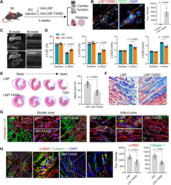
Figure 5.
Therapeutic potential of LNP–TARID in mice with myocardial infarction. (A) Schematic study design. Immediately after left anterior descending artery ligation, LNP–TARID in hyaluronic acid hydrogel was injected into the pericardial cavity. Empty lipid nanoparticles were used as control. Four weeks after injection, therapeutic efficacy was evaluated by functional and histological analysis. (B) Injection of LNP–TARID promoted Tcf21 expression in myocardial infarction hearts. Scale bar, 60 μm. (C) B-mode and M-mode images of echocardiography measurement of cardiac function. (D) Quantitative data of cardiac function. Data were expressed as mean ± SD, n = 6 mice for each group. (E) Masson trichrome staining was performed to show the infarct scar, and accordingly, infarct size was calculated. Quantitative data were expressed as mean ± SD, n = 6 mice for each group. (F) Comparison of the scar organization in the border zone of the infarct. Scale bar, 60 μm. (G) Immunofluorescence staining of Vimentin to show the alignment of fibroblast in the infarct. Scale bar, 60 μm. (H) Immunofluorescence staining of α-SMA and Collagen 1 expression in the scar and quantitative analysis. Scale bar, 60 μm. Data were expressed as mean ± SD, n = 6 mice for each group.

