Figure 6.
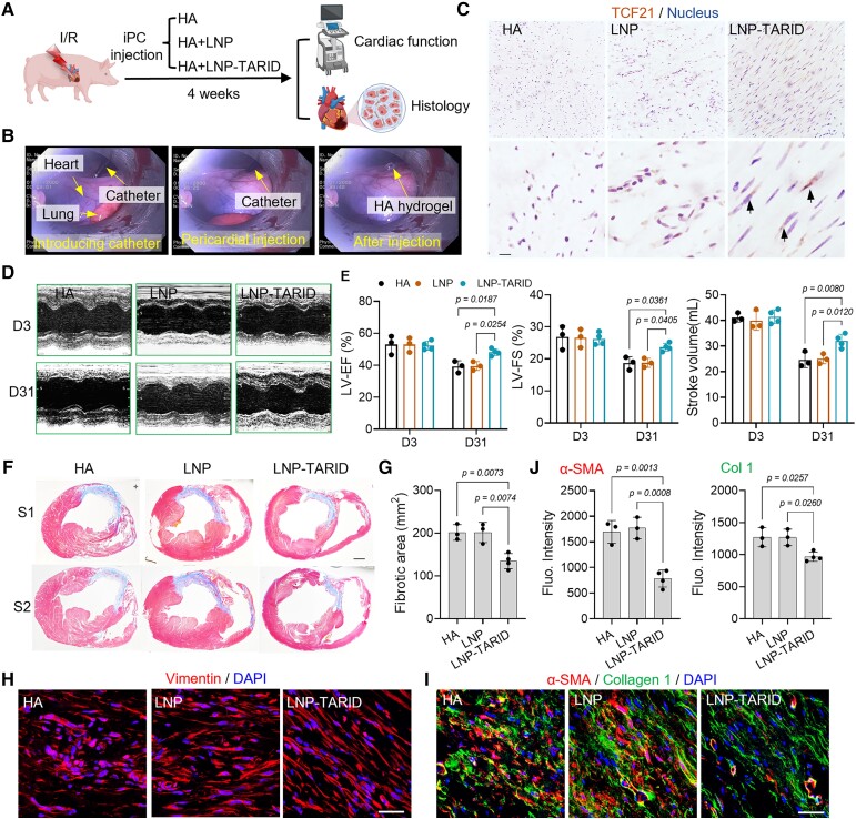
Therapeutic potential of LNP–TARID in pig ischaemia/reperfusion injury. (A) Schematic study design. Immediately after reperfusion, LNP–TARID was intrapericardially injected, and hyaluronic acid hydrogel was used as the carrier. Injection of hyaluronic acid hydrogel and empty lipid nanoparticles were used as control. Four weeks after injection, the therapeutic efficacy was evaluated by functional and histological analysis. (B) Distilled images showing minimally invasive intrapericardial injection in pigs. (C) Immunohistochemistry staining of Tcf21 in the heart. Scale bar, 100 μm (D) M-mode images of echocardiographic measurement of cardiac function. (E) Quantitative data to show the left ventricular ejection fraction, left ventricular fraction shortening, and stroke volume changes after LNP–TARID treatment. Data were acquired from three continuous cardiac cycles and expressed as mean ± SD, n = 3 pigs in HA and empty LNP treated groups; n = 4 pigs in LNP–TARID treated group. (F) Masson trichrome staining was performed to show the infarct scar, and accordingly, the fibrotic area (G) was measured. Scale bar, 1 cm. Quantitative data were expressed as mean ± SD, n = 3 pigs in hyaluronic acid and empty lipid nanoparticle–treated groups; n = 4 pigs in LNP–TARID treated group. (H) Immunofluorescence staining of Vimentin to show the fibroblasts alignment. Scale bar, 60 μm. (I) Immunofluorescence staining to detect α-SMA and Collagen 1 expression in the scar and the quantitative data (J). Scale bar, 60 μm. Quantitative data were expressed as mean ± SD, n = 3 pigs in hyaluronic acid and empty lipid nanoparticle–treated groups; n = 4 pigs in LNP–TARID treated group.

