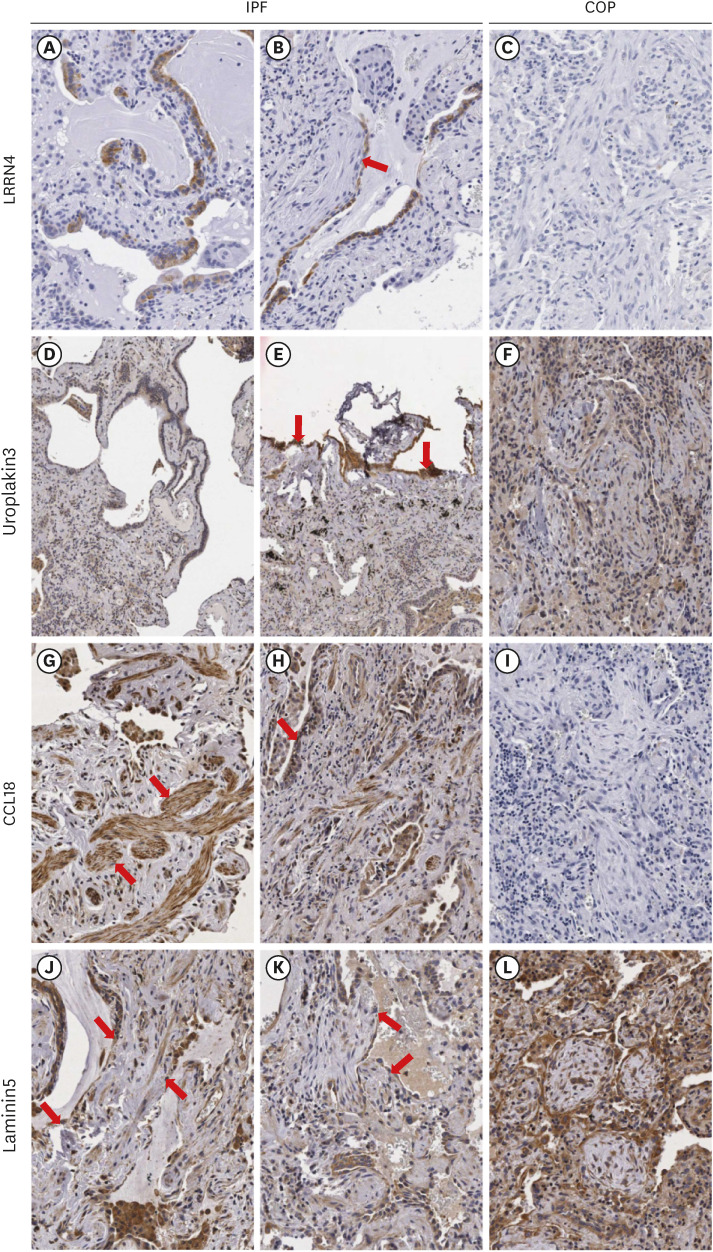
Fig. 2. Comparison of immunohistochemical staining results for LRRN 4, UPK3B, CCL18, and laminin-5 in between IPF and COP samples. LRRN4: In IPF, positive expression in the epithelial cells along the honeycombing (A) and cells covering the fibroblastic foci (B, arrow). In COP, no expression in the epithelial cells surrounding fibrous tufts (C). UPK3B: In IPF, mild expression in the epithelial cells covering honeycombings (D) and positive expression in the pleural mesothelial cells (E, arrows). In COP, weak expression in the fibrous tufts (F). CCL18: In IPF, positive expression in the smooth muscle bundles (G, arrows), positive expression in the alveolar epithelial cells (arrow), and alveolar macrophages (arrow heads) in fibrotic lesion (H). In COP, no expression in the fibrotic area (I). Laminin-5: In IPF, focal expression in honeycombing (J, arrows) and in the epithelial cells covering fibroblastic foci (K, arrows). In COP, diffuse expression in the epithelial cells surrounding the fibrous tufts (L). LRRN4 and UPK3B were clearly stained in the visceral pleura both IPF and COP samples. All markers were no or scanty staining in normal epithelium. Original magnification: A-C and F-L (×100); D and E (×40).
LRRN4 = leucine rich repeat neuronal 4, UPK3B = uroplakin 3B, CCL18 = CC-chemokine ligand 18, IPF = idiopathic pulmonary fibrosis, COP = cryptogenic organizing pneumonia.

