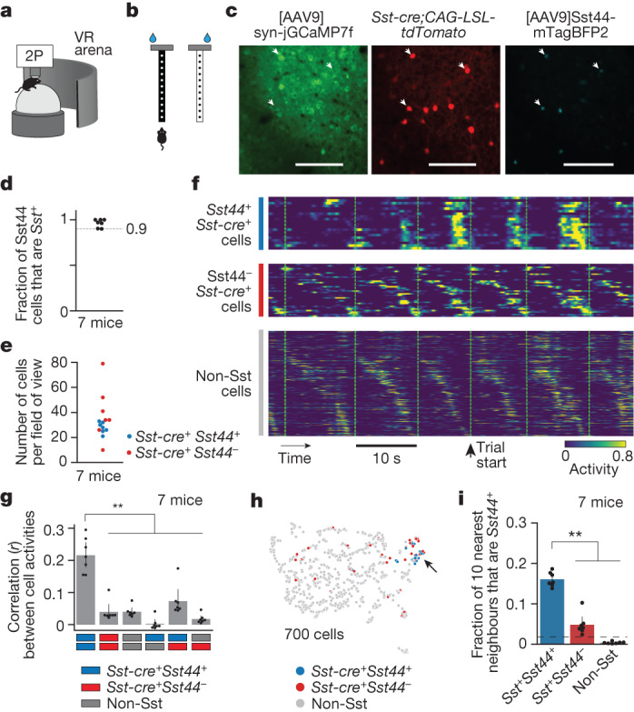
Fig. 2. Sst44 cells activate synchronously in a cell-type-specific pattern.

a, Imaging activity during virtual navigation. 2P, two-photon scanning microscope; VR, virtual reality. b, T-maze virtual environments. c, Example cropped field of view. Scale bars, 100 μm. d, The specificity of the Sst44 enhancer. e, The number of Sst cells per field of view across three planes. f, Sample activity trace showing synchronous activity in Sst44 neurons. Each row is the activity of one cell. g, Pearson correlation between cells of each cell type. h, UMAP projection of each cell’s activity from one session, showing clustering of Sst44 neurons. i, The fraction of the ten nearest neighbours in activity space that are Sst44-positive. The dashed line shows the mean after shuffling cell type identities. Activity was smoothed with a 0.25 s Gaussian filter. Statistical analysis was performed using Kolmogorov–Smirnov tests. Data are mean ± bootstrapped 95% confidence intervals.
