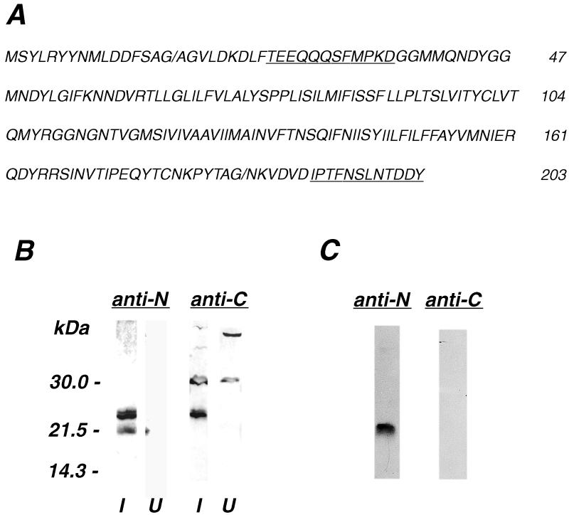
FIG. 1.
C-terminal cleavage of the A17L protein. (A) The A17L ORF is shown with the sequences used to generate synthetic peptides for immunization underlined and a slash at the known AGA and predicted AGN cleavage motifs. The antibodies induced by peptides TEEQQQSFMPKD and IPTFNSLNTDDY are referred to as A17L N antibody and A17L C antibody, respectively. (B) Western blot of extract from uninfected cells (U) and cells infected with vaccinia virus (I) and probed with A17L N antibody (anti-N) and A17L C-antibody (anti-C). The masses and positions of marker proteins are indicated on the left. Close inspection reveals that the 23- to 25-kDa bands from infected cells detected with N and C antibodies are doublets. The C antibody cross-reacted with more slowly migrating bands from uninfected and infected cells. (C) Western blot of proteins from 11 μg of sucrose gradient-purified vaccinia virions probed with A17L N and C antibodies.

