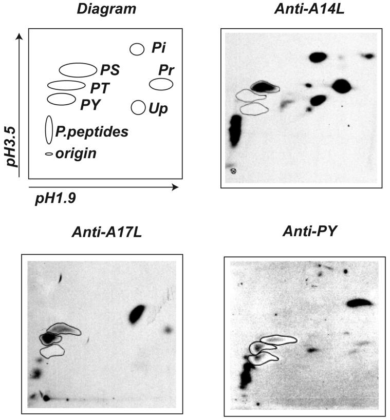
FIG. 9.
Phosphoamino acid analysis. BS-C-1 cells were infected with vaccinia virus vA17LΔ5 in the presence of IPTG and labeled overnight with 32Pi. After lysis and immunoprecipitation with A14L antibody, A17L N antibody, or antibody to phosphotyrosine (anti-PY), the proteins were resolved by SDS-PAGE and transferred to a PVDF membrane. The radioactively labeled bands were excised, hydrolyzed with HCl, and analyzed by two-dimensional thin-layer electrophoresis at pH 1.9 and 3.5. The standards amino acids were visualized with ninhydrin, and the plates were autoradiographed. The spots corresponding to phosphoserine (PS), phosphothreonine (PT), phosphotyrosine (PY), Pi, phosphoribose (Pr), and phosphouridine (Up) are identified. P.peptides, phosphorylated peptides.

