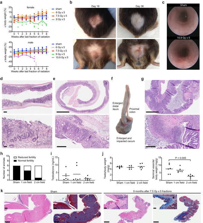
Fig. 3. Acute dose-limiting toxicity and late effects of hypofractionated stereotactic ablative radiotherapy (SART) to the pelvis.
a Body weight change relative to pretreatment baseline for female and male mice treated with 6–10.9 Gy x 5 fractions QOD. Individual animals are shown with group means connected. Female n = 6 for 0 and 7.5 Gy, n = 3 for 6 and 9 Gy. Male n = 9 for 0, 7.5, and 9 Gy, n = 3 for 6 and 10.9 Gy. Each asterisk (*) indicates time of death or protocol mandated euthanasia of a single animal. b Representative skin changes in a male mouse at the indicated time points after completing 10.9 Gy x 5 fractions QOD to a 2 cm pelvic field. Upper panels show dorsal side, lower panels ventral side. c Endoscopic images show colorectal mucosa in an untreated animal and 1 week after completing 10.9 Gy x 5 fractions QOD to a 2 cm pelvic field. d H&E shows loss of mucosal integrity in the rectum 5.5 weeks after completing 10.9 Gy x 5 fractions QOD to a 2 cm pelvic radiation field. Scale bar = 250 µm for the low power view and 100 µm for the high-power view. e H&E shows severe chronic proctitis 4 months after completing 14.4 Gy x 3 fractions evenly distributed over 9 days to a 2 cm pelvic radiation field. Scale bar = 2 mm for the low power view and 300 µm for the high-power view. f Gross pathology of intestines 11 weeks after completing 9 Gy x 5 fractions QOD to a 2 cm pelvic radiation field. g H&E shows ulceration and inflammation in the cecum 11 weeks after completing 9 Gy x 5 fractions QOD to a 2 cm pelvic radiation field. Corresponds to the gross specimen shown in (f). Scale bar = 2 mm for the low power view and 300 µm for the high-power view. h Fertility of CD-1 male mice 9 months after sham treatment or 7.5 Gy x 5 fractions QOD to a 1 or 2 cm pelvic radiation field. Normal fertility is defined as ability to father a litter while housed with a female of breeding age for one month. i Serum testosterone from animals in (h). j Wet weight of testis and seminal vesicles from animals in panel h. P value estimate by unpaired, two-tailed t test. k Representative sagittal section of one seminal vesicle in CD-1 male mice 8.5 months after sham treatment or 7.5 Gy x 5 fractions QOD to a 2 cm pelvic field. H&E and blue trichrome stain showing fibrosis (black arrowhead). Note that secretions in the seminal vesicle lumen also stain blue. Scale bar = 1 mm. Inset shows higher magnification of the seminal vesicle wall. Scale bar = 100 µm. All 5-fraction regimens were delivered over a period of 9 days as shown in Table 1.

