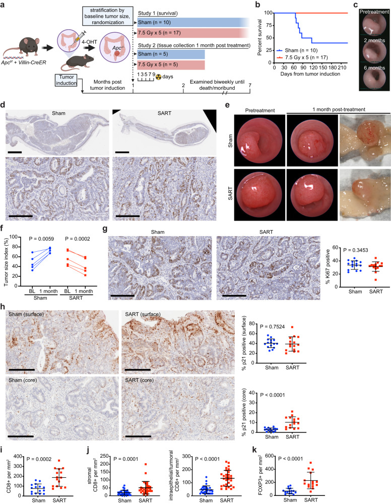
Fig. 7. Stereotactic ablative radiotherapy (SART) improves survival in an autochthonous model of colorectal adenoma.
a Graphical representation of colorectal cancer GEMM and study design, including timing of interventions. Study 1 was conducted with approximately equal numbers of male and female mice that were 2–3 months of age at time of tumor induction. Study 2 involved only male mice. 4-OHT, 4-hydroxytamoxifen. b Overall survival in study 1. c Representative endoscopic images of rectal adenoma prior to treatment and at the indicated time points after SART. d Representative Ki67 IHC of a rectal adenoma 7 months after radiation (8 months after tumor induction). A tumor from an untreated animal, two and a half months after tumor induction, is shown for comparison. Scale bar = 2 mm (low power view), 0.2 mm (high power view). e Representative endoscopic and gross images of a SART-treated tumor and untreated control from study 2. f Size of individual tumors from study 2 as evaluated by endoscopy at baseline (BL) and 1-month post SART. n = 5 for both treatment groups. P value estimate by paired, two-tailed t test. g Representative Ki67 IHC images and proliferative index of tumors from study 2. Three high power fields per tumor were assessed. Scale bar = 0.2 mm. P value estimate by Mann–Whitney test. h Representative p21 IHC images and quantitation of p21 positive cells at the surface and core region of tumors from study 2. Three high power fields per region/tumor were assessed. Scale bar = 0.2 mm. P value estimate by unpaired, two-tailed t test. i Quantitation of cytotoxic T cells assessed by IHC in tumors from Study 2. Three high power fields per tumor were assessed. P value estimate by unpaired, two-tailed t test. j Quantitation of cytotoxic T cells assessed by IHC in stromal vs intraepithelial/tumor compartments of tumors from Study 2. Six high power fields per tumor were assessed. P value estimate by unpaired, two-tailed t test. k Quantitation of regulatory T cells assessed by IHC in tumors from Study 2. Three high power fields per tumor were assessed. P value estimate by unpaired, two-tailed t test.

