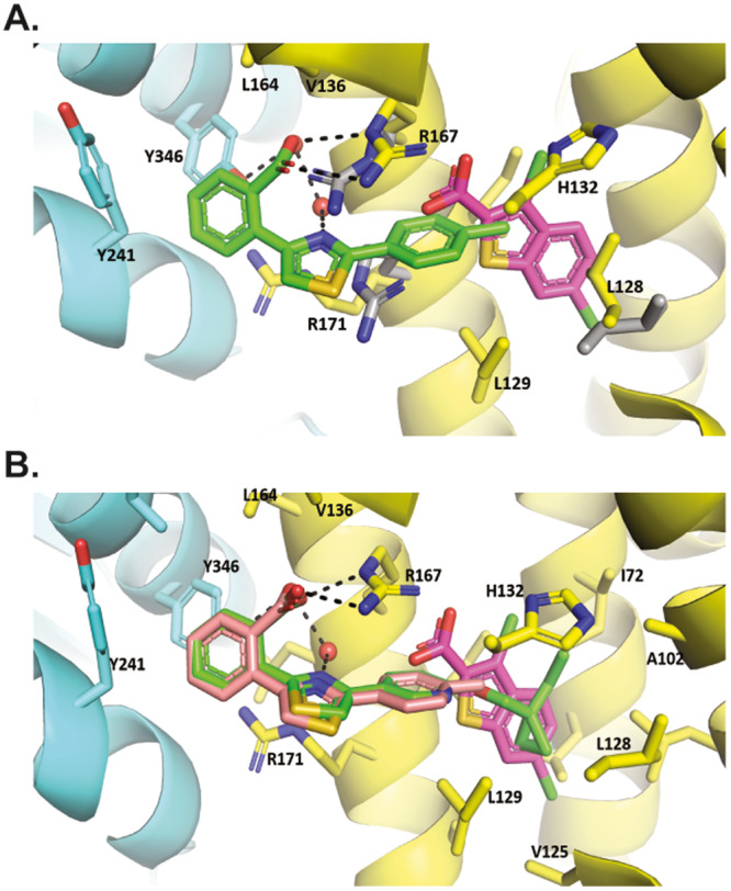
Fig. 3. Thiazoles bind to BDK in distinct mode adjacent to thiophenes.

Allosteric binding site from X-ray crystal structures of BDK bound to inhibitors. The regulatory domain of BDK is shown in yellow ribbons, and the kinase domain in aqua. Side chains of contacting residues are shown, and hydrogen bonds in dash. A S3 (stick model, C atoms colored in green) bound to hBDK. Superimposed is the BT2 X-ray crystal structure (PDB ID: 4E00) with BT2 C atoms colored in pink and side chains of residues that adopt different rotamers in the BT2 complex in gray. B PF-07247685 (stick model, C atoms colored in green) bound to hBDK. Superimposed are BT2 (C atoms colored in pink) and S3 (C atoms colored in salmon).
