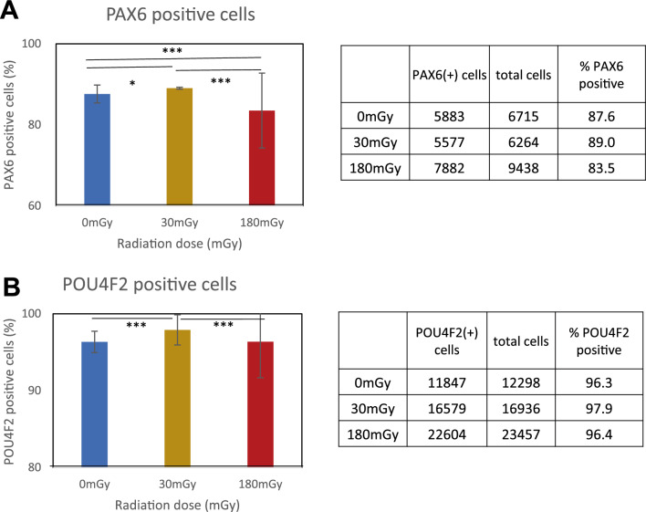
Figure 5.
Immunofluorescence of PAX6 and POU4F2 on day 35 after 30 or 180 mGy of irradiation from day 4 to 5. Immunofluorescence with anti-PAX6 or anti-POU4F2 antibody and beta-tubulin were stained. DAPI was analyzed with IN CELL Analyzer. Nuclear area was identified with fluorescence intensity and area size of DAPI. Then, fluorescence intensity of the target in the nuclear area was analyzed. (A) PAX6-positive cells. (B) POU4F2-positive cells. *P < 0.05, ***P < 0.001 (chi-squared test) (n = 3). Experiment was repeated three times and representative data was presented.

