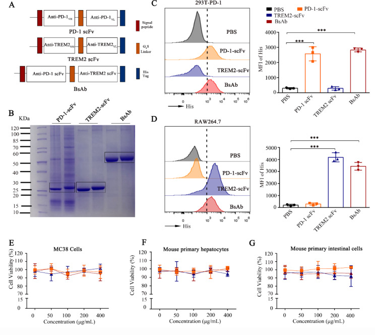
Fig. 2.
Construction and characterization of the bi-specific scFv antibody (BsAb). (A) Schematic representation of PD-1-scFv, TREM2-scFv, and BsAb. (B) SDS-PAGE analysis of PD-1 scFv, TREM2 scFv, and BsAb derived from the supernatants of CHO cells using nickel column purification and concentration. (C) Flow cytometric histograms demonstrating His-tag detection of PD-1 scFv, TREM2 scFv, and BsAb binding to 293T stably expressing PD-1 (293T-PD-1). The 293T-PD-1 cells were treated with PD-1 scFv, TREM2 scFv, and BsAb at 200 µg/mL and MFI of His was detected by flow cytometry. Bar plots summarizing the data are shown on the right. (D) Flow cytometric histograms demonstrating His-tag detection of PD-1 scFv, TREM2 scFv, and BsAb binding to TREM2 on the RAW 264.7 cells. PD-1 scFv, TREM2 scFv, and BsAb at 200 µg/mL and MFI of His was detected by flow cytometry. Bar plots summarizing the data are shown on the right. (E-F) Relative viability of MC38 cells, mouse primary hepatocytes, and mouse primary intestinal cells treated with PD-1 scFv, TREM2 scFv, and BsAb at 200 µg/mL for 24 h analyzed using the CCK-8 kit. Data are expressed as means ± SD; * P < 0.05, **P < 0.01 (n = 3), by unpaired Student’s t test (C-D). PD-1, programmed death-1; scFv, single-chain fragment variable; TREM2, triggering-receptor-expressed on myeloid cells 2; SDS-PAGE, sodium dodecyl-sulfate polyacrylamide gel electrophoresis; CHO, Chinese hamster ovary; RAW 264.7, Mouse Mononuclear Macrophages Cells; 293T, human embryonic kidneys; CCK, cell counting kit; SD, standard deviation

