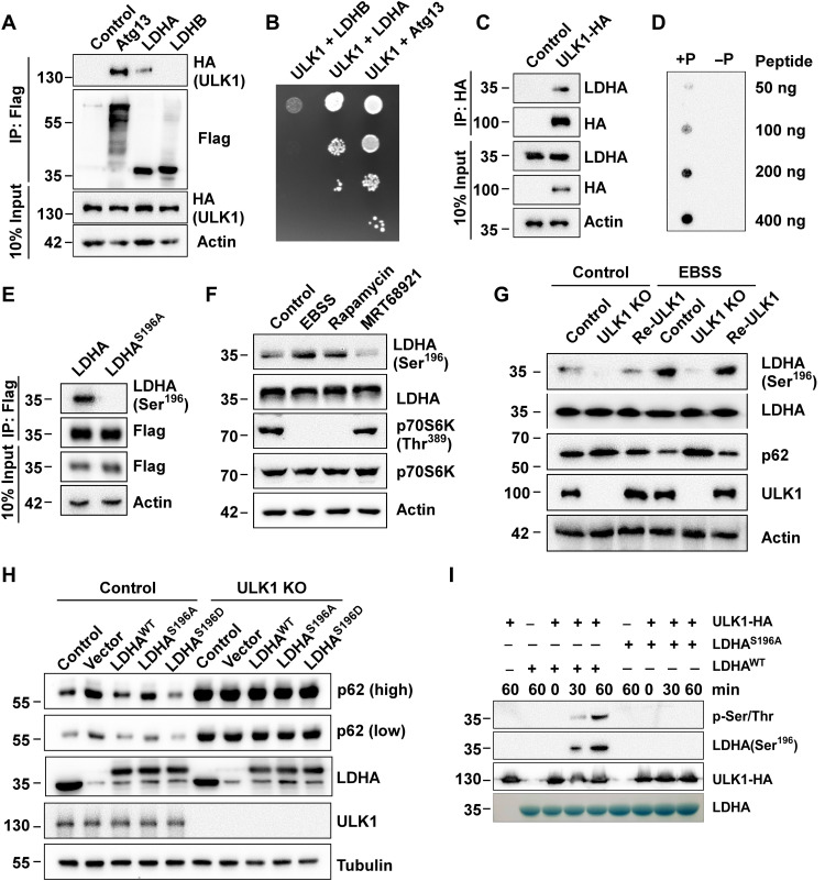
Fig. 1. ULK1 induces LDHA Ser196 phosphorylation.
(A) ULK1 interacts with LDHA. Flag-tagged Atg13, LDHA, or LDHB were coexpressed with HA-ULK1 individually, and immunoprecipitation was performed using anti-Flag beads. Samples were analyzed by anti-HA Western blot. (B) Interaction assay of ULK1 and LDHA by yeast two-hybrid assay. pGBKT7-Atg13, pGBKT7-LDHA, or pGBKT7-LDHB and pGADT7-ULK1 were cotransformed into the yeast strain MJ109 and grown on the agar plate without leucine and tryptophan (YPD/−Leu/−Trp). Atg13 as positive control. (C) Interaction assay of ULK1 and endogenous LDHA. ULK1-HA was expressed in HEK293T cells. Immunoprecipitation was performed using anti-HA beads and analyzed by Western blot with antibody to LDHA. (D) Antibody specificity was analyzed by dot blot assay with a LDHA Ser196 phosphorylation antibody. +P peptide represent phosphorylation modification of LDHA Ser196. −P peptide represent nonphosphorylation modification of LDHA Ser196. (E) Antibody specificity test. LDHAWT-Flag or LDHAS196A-Flag was expressed in HEK293T cells. Immunoprecipitation was performed by anti-Flag beads, and LDHA Ser196 phosphorylation levels were analyzed by Western blot with phospho-LDHA Ser196 antibody. (F) LDHA Ser196 phosphorylation assay. HEK293T cells were treated with EBSS medium, mTOR inhibitor rapamycin, and ULK1 inhibitor MRT68921 for 4 hours. LDHA Ser196 phosphorylation was analyzed by phospho-LDHA Ser196 antibody. (G) LDHA Ser196 phosphorylation assay in ULK1-KO MEF cells. MEF cells and ULK1-KO MEF cells were transfected with vector or ULK1-HA and were cultured in complete medium or EBSS medium for 3 hours. LDHA Ser196 phosphorylation was analyzed by phospho-LDHA Ser196 antibody. (H) Autophagy assay in LDHA knockdown control cells and ULK1-KO cell (followed by expression of either LDHAWT, LDHAS196A, or LDHAS196D). (I) LDHA Ser196 phosphorylation assay in vitro. ULK1 was purified by immunoprecipitation from HEK293T cells, and its kinase activity in vitro was analyzed using purified LDHAWT-Flag and LDHAS196A-Flag from E. coli as substrate. LDHA Ser196 phosphorylation was analyzed by anti–phospho-LDHA Ser196 and anti–phospho-Ser/Thr antibody.

