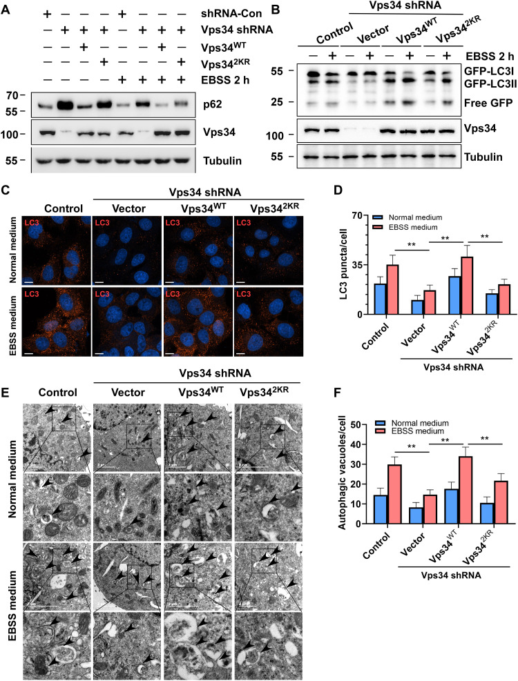
Fig. 5. Vps34 lactylation facilitates autophagosome formation and maturation.
(A) p62 degradation assay. HEK293T cells (control shRNA and Vps34 KD/rescued by vector, Vps34WT and Vps342KR) were treated with normal medium or EBSS medium for 2 hours. p62 was analyzed by Western blot. (B) GFP-LC3 cleavage assay. HEK293T cells (control shRNA and Vps34 KD/rescued by vector, Vps34WT, and Vps342KR) were treated with normal medium or EBSS medium for 2 hours and analyzed by Western blot with GFP antibody. (C) LC3 puncta assay. U2OS cells (control shRNA and Vps34 KD/rescued by vector, Vps34WT, and Vps342KR) were treated with normal medium or EBSS medium for 2 hours. LC3 puncta was analyzed by confocal microscopy. Scale bar, 10 μm. (D) Quantification of LC3 puncta. Data are shown as means ± SD; **P < 0.01, n = 30. (E) Autophagic vacuole assay. U2OS cells (control shRNA and Vps34 KD/rescued by vector, Vps34WT, and Vps342KR) were treated with normal medium or EBSS medium for 2 hours. Autophagic vacuole was analyzed by transmission electron microscopy. (F) Quantification of autophagic vacuole. Data are shown as means ± SD; **P < 0.01, n = 20.

