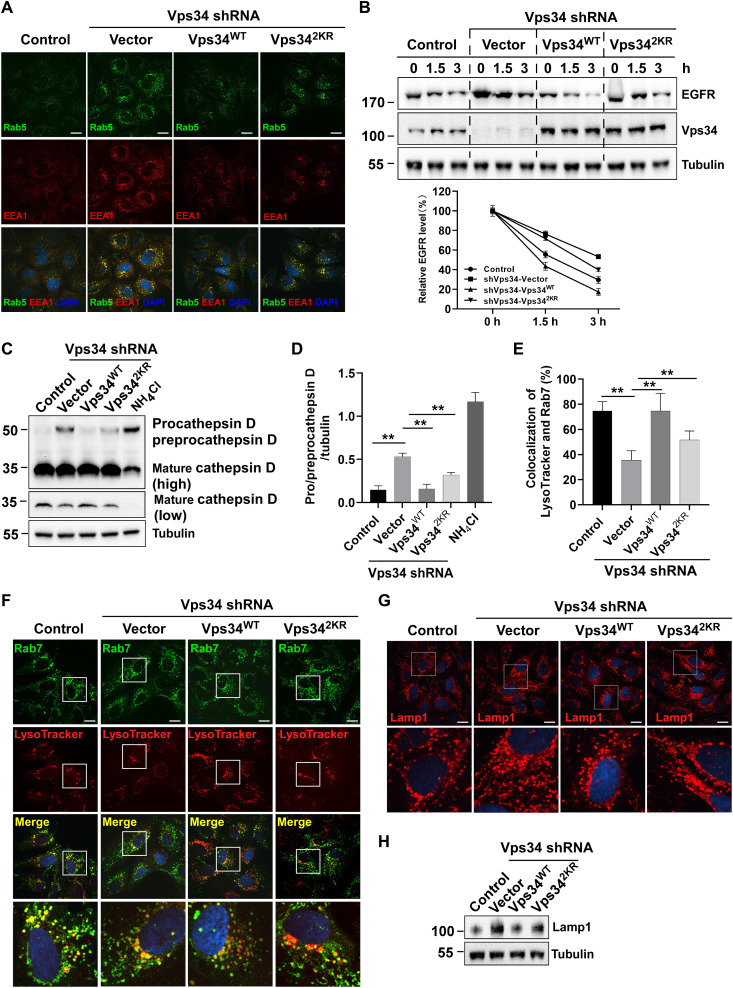
Fig. 6. Vps34 lactylation promotes endosome-lysosomal degradation.
(A) The Rab5 and EEA1 localization in U2OS cells (control shRNA and Vps34 KD/rescued by vector, Vps34WT, and Vps342KR). Rab5 and EEA1 colocalized on enlarged endosomes in Vps34 delactylation cells. Scale bar, 10 μm. (B) EGFR degradation assay. HEK293T cells (control shRNA and Vps34 KD/rescued by vector, Vps34WT, and Vps342KR) were treated with EGF (100 ng/ml) for 0,1.5, and 3 hours. EGFR were analyzed by Western blot. EGFR levels were quantified by ImageJ software and normalized by percentage of 0-hour EGFR level. (C) Cathepsin D maturation assay in U2OS cells (control shRNA and Vps34 KD/rescued by vector, Vps34WT, and Vps342KR) was analyzed by Western blot with Cathepsin D to show procathepsin D, preprocathepsin, and mature cathepsin D. (D) The levels of procathepsin D and preprocathepsin D were quantified by ImageJ software. Data are shown as means ± SD; **P < 0.01, n = 3. (E) Percentage of Rab7 and LysoTracker colocalization. Data are shown as means ± SD; **P < 0.01, n = 30. (F) Rab7 and LysoTracker colocalization in U2OS cells (control shRNA and Vps34 KD/rescued by vector, Vps34WT, and Vps342KR) was analyzed by confocal microscopy. Scale bar, 10 μm. (G) Lysosome staining assay by confocal microscopy with Lamp1 antibody in U2OS cells (control shRNA and Vps34 KD/rescued by vector, Vps34WT, and Vps342KR). Scale bar, 10 μm. (H). Lamp1 expression in U2OS cells (control shRNA and Vps34 KD/rescued by vector, Vps34WT, and Vps342KR) was analyzed by Western blot.

