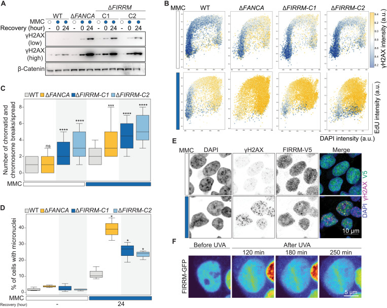
Fig. 2. FIRRM loss causes increased levels of DNA damage and genome instability.
(A) Wild-type, FANCA, or FIRRM knockout HAP1 cells were treated with 50 nM MMC for 24 hours and left to recover for the indicated time points. Whole-cell extracts were subjected to immunoblot analysis using γH2AX antibodies. β-Catenin was used as a loading control. (B) Wild-type, ΔFANCA, or ΔFIRRM HAP1 cells were left untreated or exposed to 50 nM MMC for 24 hours, allowed to recover for 24 hours; stained with γH2AX, EdU, and (DAPI); and subjected to flow cytometry analysis. n = 3. (C) Quantification of chromosome and chromatid breaks per metaphase spread (means ± SEM); 60 metaphases of KP cells left untreated or treated with 50 nM MMC for 24 hours were analyzed for each condition and pooled from three independent experiments. (D) Quantification of micronuclei in indicated genotypes, unchallenged or upon 50 nM MMC treatment and 24 hours of recovery. n > 300 cells per condition. n = 3. (C and D) Significance was calculated by a Mann-Whitney U test. ns, not significant. *P < 0.05, ***P < 0.001, and ****P < 0.0001. (E) Immunofluorescence staining for γH2AX and FIRRM-V5 in HAP1 cells upon MMC treatment (50 nM, 24 hours) or left untreated. To visualize chromatin bound protein, cells were pre-extracted before fixation. n = 3. (F) Recruitment of GFP-FIRRM to laser UVA microirradiation sites after psoralen treatment in HAP1 cells. n = 2.

