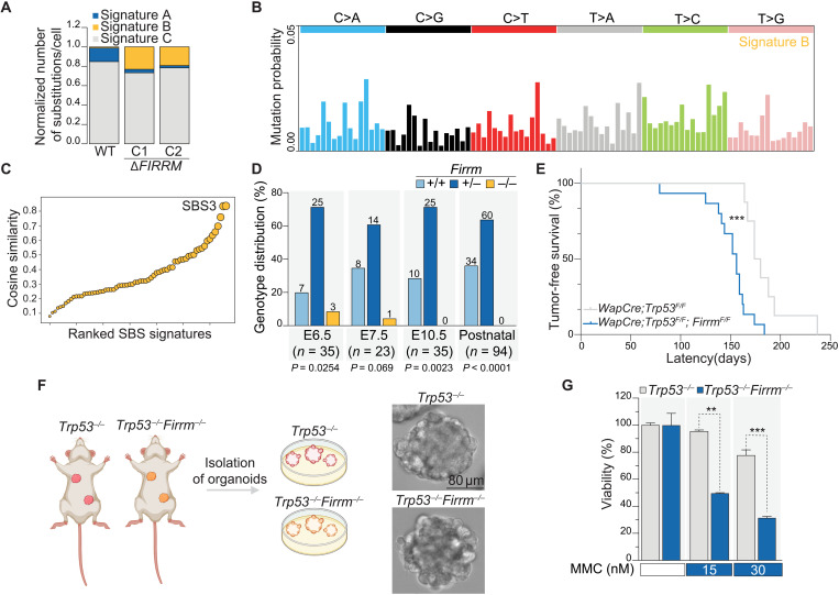
Fig. 3. FIRRM loss causes early embryonic lethality and accelerates tumorigenesis.
(A) Contribution of signatures A, B, and C in untreated wild-type and ΔFIRRM HAP1 cells. (B) Signature decomposition of wild-type and ΔFIRRM HAP1 cells in untreated conditions. (C) Cosine similarity comparing signature B to existing single base substitution (SBS) signatures. (D) Recovery of Firrm+/+, Firrm+/−, and Firrm−/− embryos at different time points after performing timed matings. Numbers of analyzed embryos are indicated, and a Chi-square test was used to determine significance. (E) Kaplan-Meier survival curve depicting the mammary tumor-free survival of WapCre;Trp53F/F (n = 8) and WapCre;Trp53F/F;FirrmF/F (n = 15) female mice. Significance was calculated by a Mantel-Cox test. ***P < 0.001, median survival of 177 days (WapCre;Trp53F/F) versus 156 days (WapCre;Trp53F/F;FirrmF/F). (F) Schematic depiction of organoid isolation from mammary tumor–bearing animals and example images of the derived organoids. (G) Viability of mammary tumor organoids after exposure to MMC for 7 days. Values were normalized to untreated conditions. n = 2. P values were calculated using a two-tailed t test. **P < 0.01 and ***P < 0.001.

