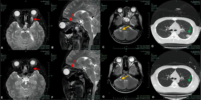
Fig. 2.
Orbital MRI, head MRI, and lung CT of case 2 (A, B, C, and D) were examined on early admission and (E, F, G, and H) were re-examined 50 days later before discharge. (D) Lung CT showing bilateral lung inflammation, which was more severe on the left side (green arrows). (C) Head MRI (T2, coronal) showing brain abscess formation (yellow arrows). (A,B) Orbit MRI (T2, coronal; T2, sagittal) showing bilateral orbital inflammation and abscess formation in the upper left orbit (red arrows). (E, F, G, and H) Follow-up MRIs and CTs showing that the inflammation and abscesses in the lungs, cerebrum, and orbits had mostly resolved before discharge

