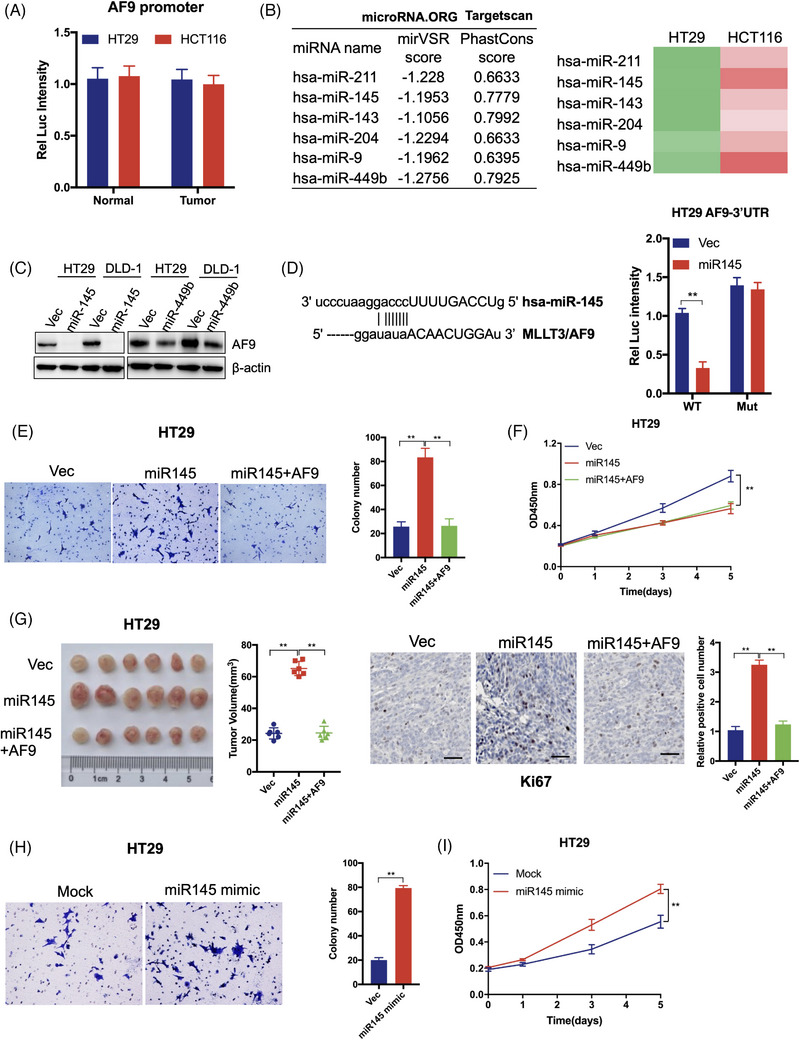
FIGURE 4.

miR‐145 targets AF9 3′ untranslated region (3′UTR) and silences AF9 mRNA. (A) Dual‐luciferase reporter assay was performed to test the AF9 promoter activity. (B) microRNA.ORG and Targetscan were used to predict the potential miRNA, which could target 3′UTR of homo sapiens AF9. The green represents low expression of miRNAs, while the red represents high expression of miRNAs. And the deeper the colour is, the higher or lower expression of miRNAs is. (C) miR145 or miR449b was transfected into HT29 or DLD‐1 cells. Western blot analysis was used to detect AF9 protein level by indicated antibodies. (D) Dual‐luciferase reporter assay was performed to test the activity of wild type or mutant 3′UTR by co‐transfection with miR‐145. E, Representative images of transwell assays performed in HT29 cells (including Vec, miR‐145 or miR‐145+AF9) are shown (scale bar represents 60 μm). (F) Cell proliferation of HT29 cells (including Vec, miR‐145 or miR‐145+AF9) was measured by CCK‐8. (G) Xenograft formation. HT29 cells (including Vec, miR‐145 or miR‐145+AF9) were implanted into left groin of nude mice (n = 6 for each group). The tumour volume was measured at the end of the experiment. Ki67 staining was performed to test the in vivo proliferation. (H) Representative images of transwell assays performed in HT29 cells transfected with or without miR‐145 mimic are shown (scale bar represents 60 μm). (I) Cell proliferation of HT29 cells transfected with or without miR‐145 mimic was measured by CCK‐8 (**p < .01).
