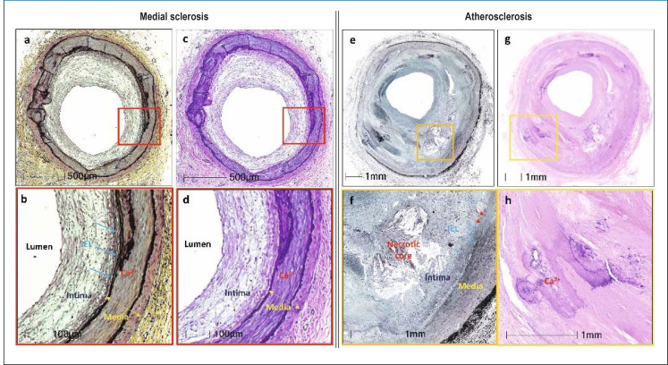
eFigure 1.
Comparison of typical histological features of medial sclerosis (a–d) and atherosclerosis (e–h)
Both diseases are shown in lower (a, b and e, f, respectively) and higher (c, d and g, h, respectively) magnification.
The typical histological picture of medial sclerosis of the popliteal artery is depicted. Medial sclerosis is characterized by circular calcification within the media and moderate intimal hyperplasia. The typical histological picture of atherosclerosis of the superficial femoral artery is shown. Intimal lesion with necrotic nucleus, penetration of the internal elastic lamina and calcified focus, clearly assigned to the intima, are shown. Internal elastic lamina (IEL) marked with arrows.
Adopted from Lanzer et al. Eur Heart J 2022; 43: 2824–6.

