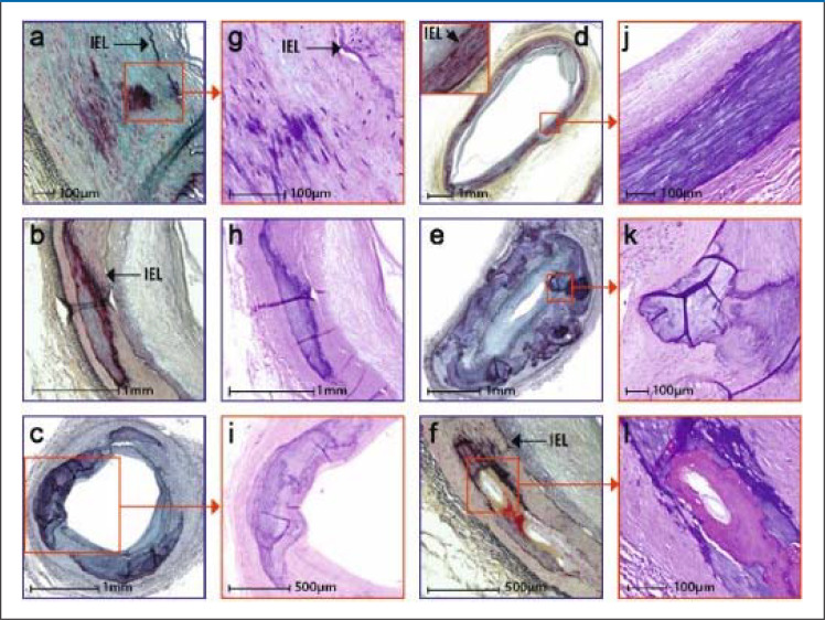
eFigure 2.
Histological stages and histological characteristics of medial sclerosis
Staging is based on the extent of calcifications.
The various stages and morphological characteristics are shown in lower (Figures a, b, c, d, e, f) and higher (Figures g, h, i, j, k, l) magnifications.
Stage I: Calcifications of the elastica interna with or without extension into the media (Figures a, g).
Stage II: Formation of larger calcifications (1 to 3 mm) (Figures b, h)
Stage III: Calcifications >3 mm to >90° of medial circumference (Figures c, i)
Stage IV: Annular calcifications of the entire circumference (Figures d, j)
Nodular calcifications as globular calcification foci with fibrin deposits (Figures e, k)
nd bone formation as proper bone foci, rarely associated with chondroid metaplasia, occur mostly in stages III/IV, rarely in stage II (Figures f, l).
Lanzer et al. J Am Coll Cardiol 2021; 78:1145–65 by courtesy of Elsevier)

