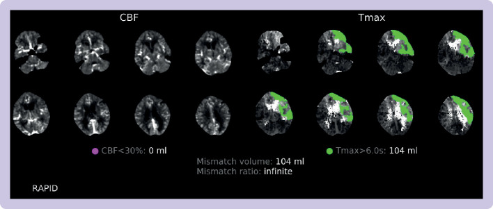
Figure 6.
Computed tomography perfusion and radiological mismatch, acquired from the Rapid.AI software package, for a patient presenting to Imperial College Healthcare Trust in the late window (ie, 6-24 hours from symptom onset or last known being well). The DAWN and DEFUSE-3 trials9 10 defined core infarct as volume of brain tissue with cerebral blood flow (CBF) <30% (highlighted on the sequences on the left) and penumbra as the mismatch in volume between the hypoperfusion lesion (ie, the volume of brain tissue where it takes >6 seconds for the contrast bolus to reach maximal density (Tmax), seen on the sequences on the right). Key values at the bottom of the figure are the core infarct volume (which must be <70 mL), mismatch ratio (which must be >1.8), and penumbra or mismatch volume (which must be >15 mL)

