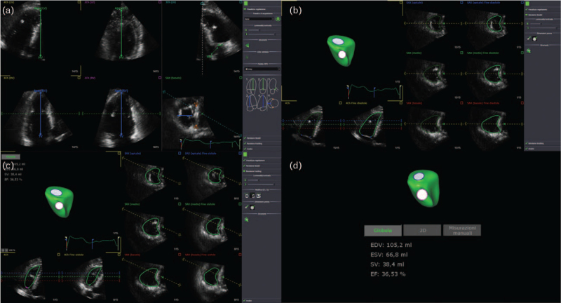
Fig. 1.
Phases of the postprocessing of echocardiographic images with GE EchoPAC software. (a) Finding landmarks with 4D RV function. (b) ‘Beutel revision phase’: the endocardial edge detection of the RV in the end-diastolic phase. (c) ‘Tracking revision phase’: the intracavitary perimeter was identified by the software during the entire cardiac cycle. (d) RV parameters and 3-D reconstruction. RV, right ventricle.

