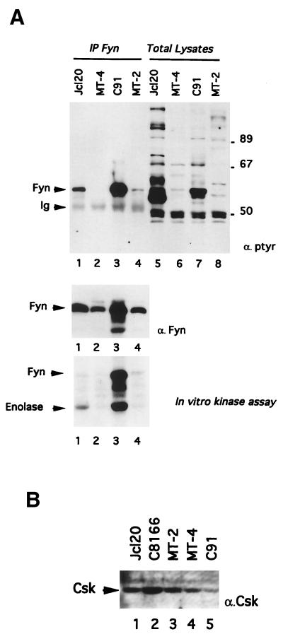
FIG. 3.
Enhanced tyrosine phosphorylation and kinase activity of Fyn parallels the decreased expression of Csk in an HTLV-1-infected T-cell line, C91. (A) After cell lysis, Fyn was immunoprecipitated and probed by immunoblotting with either antiphosphotyrosine (top panel, lanes 1 to 4) or anti-Fyn (middle panel) antibodies. Lanes 5 to 8 represent total cell lysates. The bottom panel represents the enzymatic kinase activity of Fyn as assessed by immune-complex kinase reactions in the presence of enolase. In this figure, the positions of Fyn, immunoglobulins (Ig), and enolase are indicated by arrows on the left. (B) Direct Western blot of Csk. Lysates (50 μg of protein) from Jcl20 (lane 1), C8166 (lane 2), MT-2 (lane 3), MT-4 (lane 4), or C91 (lane 5) were subjected to SDS-PAGE and Western blotting with anti-Csk antibody.

