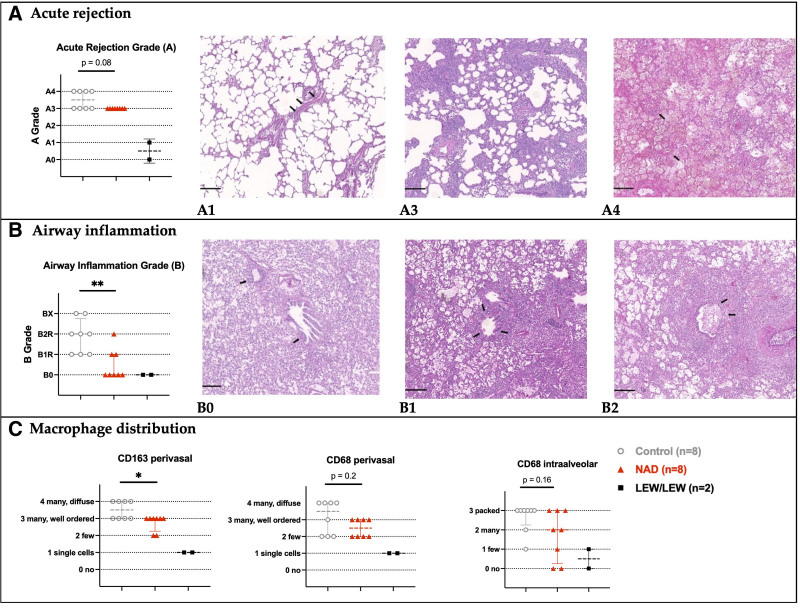
FIGURE 3.
Histological assessment at autopsy, 5 d posttransplant. A, Acute cellular rejection grades in the control, NAD+, and syngraft (LEW/LEW) groups, according to standard ISHLT guidelines. A single layer of lymphocytes around blood vessels (arrows) not visible on low magnification (A1). Prominent and easily recognizable perivascular lymphocytic infiltrate (A2, not shown since not observed). Prominent perivascular lymphocytic infiltrates with infiltration into adjacent alveolar septa (A3). Diffuse perivascular, interstitial, and alveolar inflammation and necrotizing vasculitis (arrows) (A4). B, Airway inflammation grades in the control, NAD+, and syngraft groups (LEW/LEW), according to standard ISHLT guidelines. No evidence of bronchiolar inflammation (B0). Lymphocytes in the walls of small airways (arrows) without epithelial damage (B1), intraepithelial lymphocytosis, and ulceration (arrows) of small airways (B2). C, CD163+ and CD68+ macrophage distributions in control, NAD+, and syngraft (LEW/LEW) groups based on immunohistochemistry. Notes: Histological images in hematoxylin and eosin staining, scale bars = 200 μm. The stars are significant P values vs the control. *P ≤ 0.05; **P ≤ 0.01. ISHLT, International Society of Heart and Lung Transplantation; NAD+, β-nicotinamide adenine dinucleotide.

