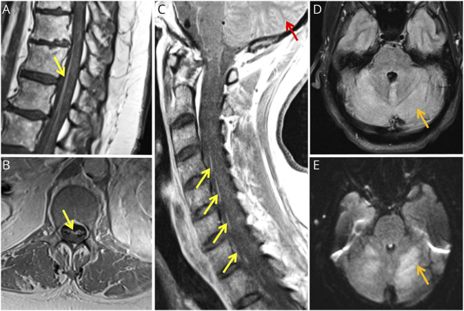
Figure 1. MR Images of Neuroinvasive WNV Manifestations in 4 Patients With MS Treated With Ocrelizumab.
MRI of the lumbosacral spine T1-weighted sequences postgadolinium infusion in case 1 in sagittal (A) and axial (B) views showing anterior nerve root enhancement (yellow arrows). (C) MRI of the cervical and thoracic spine T1-weighted sequences postgadolinium infusion in case 2 in sagittal view showing anterior nerve root enhancement (yellow arrows) and diffuse leptomeningeal enhancement of the cerebellar folia (red arrow). MRI of the brain T2-weighted fluid-attenuated inversion recovery (FLAIR) sequences in axial views in case 3 (D) and case 4 (E) showing signal hyperintensity of the cerebellum (orange arrows).

