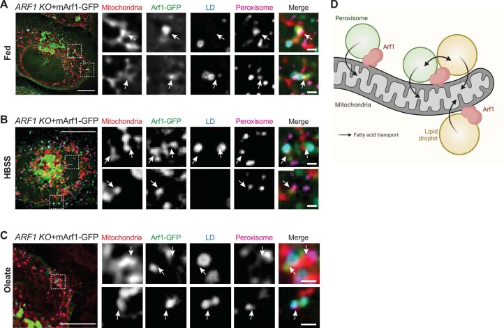
Extended Data Fig. 10. mArf1 is present at organellar contact sites.
(A–C) mArf1-GFP localization in ARF1 KO cells grown in complete media (A), shifted for 14 h in HBSS (B) or in the presence of oleate (C). Arrows indicate the presence of mArf1 at mitochondria-LD contact sites, mitochondria-peroxisomes contact sites or mitochondria-LD-peroxisomes contact sites. Scale bar: 10 µm, Scale bar inlays: 1 µm. (D) Schematic of Arf1 localization based on images taken in (A–C) and in Extended Data Fig. 9A–C. Created with Biorender.com.

