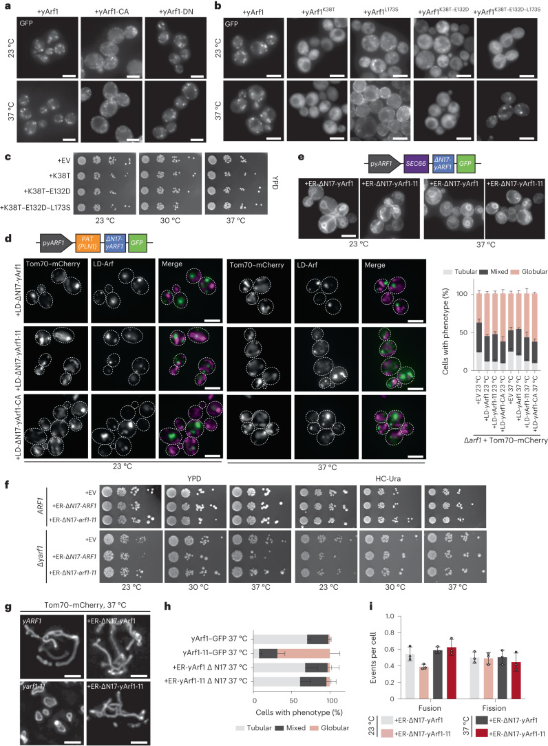
Fig. 4. LD-localized yArf1 induces mitochondria fragmentation.
a, Localization of yArf1, constitutively active (CA) or dominant negative (DN) forms of yArf1 fused to GFP grown at 23 °C or shifted to 37 °C for 30 min. Constructs were expressed from the centromeric low copy number plasmid pGFP33. Scale bar, 5 µm. b, Localization of yArf1, or yArf1 bearing single (K38T, L173S), double (K38T–E132D) or triple (K38T–E132D–L173S) substitution yarf1-11 mutations fused to GFP in Saccharomyces cerevisiae (YPH500) grown at 23 °C or shifted to 37 °C for 30 min. Constructs were expressed from the centromeric low-copy-number plasmid pGFP33. Scale bar, 5 µm. c, Growth assay of the WT strain bearing the empty pGFP3 vector (+EV), single (K38T, L173S), double (K38T–E132D) or triple (K38T–E132D–L173S) yarf1-11 mutations fused to GFP on rich YPD plates incubated at 23 °C, 30 °C or 37 °C. d, Schematic of the construct designed to anchor yArf1 on the LD via the PAT domain of the perilipin PLN1. yARF1 deleted in its myristoylation sequence (∆N17) was expressed from its endogenous promoter and fused to GFP on its 3′ end. Localization of LD-anchored ∆N17-yArf1–GFP, the constitutively active mutant yArf1-CA, or yArf1 bearing yArf1-11–GFP variant in cells depleted of ARF1 grown at 23 °C and shifted to 37 °C. Tom70–mCherry was used as a mitochondrial marker. Mitochondria phenotypes (tubular, mixed or globular) were measured. Mean and standard deviation are shown. At 23 °C, ∆yarf1 = 406 cells, ∆yarf1 + LD-Arf1 = 477 cells, ∆yarf1 + LD-Arf1-11 = 483 cells, ∆yarf1 + LD-Arf1-CA = 403 cells; At 37 °C, ∆yarf1 = 443 cells, ∆yarf1 + LD-Arf1 = 480 cells, ∆yarf1 + LD-Arf1-11 = 523 cells, ∆yarf1 + LD-Arf1-CA = 529 cells from n = 3 biological replicates. Scale bar, 5 µm. e, Schematic of the construct designed to anchor yArf1 on the ER via Sec66. yARF1 deleted in its myristoylation sequence (∆N17) was expressed from its endogenous promoter and fused to GFP on its 3′ end. Localization of ER-anchored ∆N17-yArf1–GFP or yArf1 bearing yArf1-11–GFP variant in ∆yarf1 cells grown at 23 °C and shifted to 37 °C. Scale bar, 5 µm. f, Growth assay of the ER-anchored ∆N17-yArf1–GFP or Arf1 strains bearing yarf1-11 mutations (Arf1K38T–E132D–L173S) on rich YPD plates or synthetic medium lacking uracil (HC-Ura) incubated at 23 °C, 30 °C or 37 °C, and of the ER-anchored ∆N17-yArf1–GFP or yArf1 bearing yarf1-11 mutations (Arf1K38T–E132D–L173S) in YPH500 cells lacking yARF1 (∆yarf1) on rich YPD plates or synthetic media lacking uracil (HC -Ura) incubated at 23 °C, 30 °C or 37 °C. g, Cells expressing yArf1/11 fused to GFP, or expressing ER-∆N17-yArf1/11–GFP were grown at 37 °C for 30 min and mitochondria were imaged with Tom70–mCherry by high-resolution microscopy followed by deconvolution. A z-projection of maximum intensities is shown for each panel. Scale bar, 2 µm. h,i, Measurements of mitochondria phenotypes (tubular, mixed or globular) based on images taken in g (h), and mitochondrial fusion and fission events based on Supplementary Videos 9–12 (i). Mean and standard deviation are shown. ∆yarf1 + yArf1–GFP = 364 cells, ∆yarf1 + yArf1-11–GFP = 800 cells, ∆yarf1 + ER-yArf1–GFP = 608 cells, ∆yarf1 + ER-yArf1-11–GFP = 542 cells from n = 3 biological replicates (h); at 23 °C ∆yarf1 + ER-yArf1–GFP = 210 cells, ∆yarf1 + ER-yArf1-11–GFP = 208 cells and at 37 °C ∆yarf1 + ER-yArf1–GFP = 195 cells, ∆yarf1 + ER-yArf1-11–GFP = 190 cells from n = 3 biological replicates (i). Source numerical data are available in source data.

