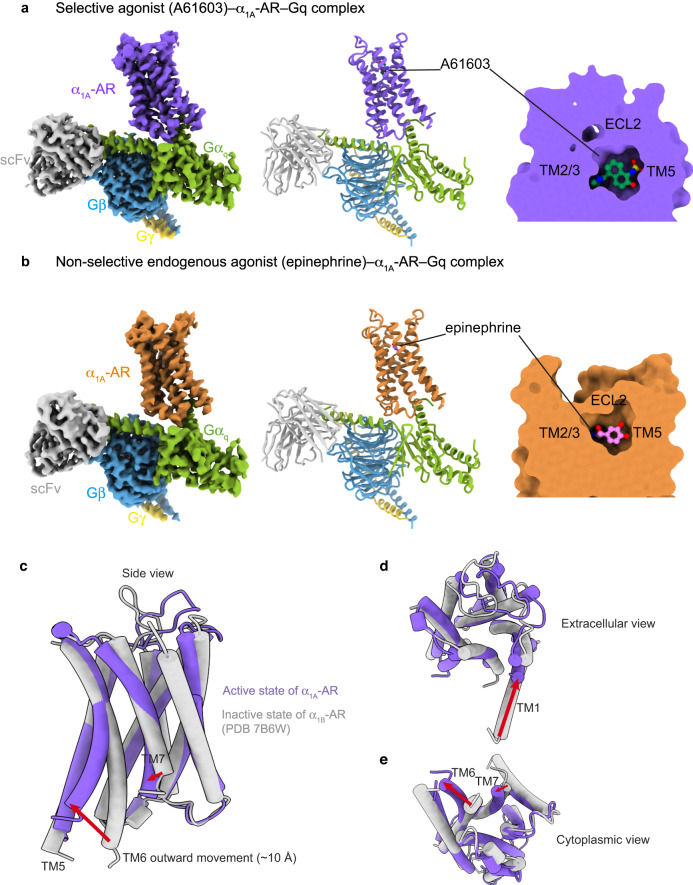
Fig. 1. Cryo-EM structures of the complexes of A61603–α1A-AR–Gq and epinephrine–α1A-AR–Gq.
a The density map, the model, and the ligand-binding pocket of A61603–α1A-AR–Gq are shown. b The density map, the model, and the ligand-binding pocket of epinephrine–α1A-AR–Gq are shown. A61603-bound α1A-AR is colored in purple. Epinephrine-bound α1A-AR is colored orange. Gαq in green. Gβ in blue. Gγ in yellow. c–e Different views of the inactive state α1B-AR (gray; PDB 7B6W) and the active state of α1A-AR in complex with Gq (this work).

