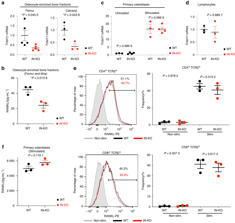
Fig. 5.
Intronic enhancer deletion decreases RANKL expression in osteocytic cells but not in other cell types. a Tnfsf11 mRNA expression levels in osteocyte-enriched femur (n = 5 per group) and calvaria (n = 3 per group) bone fractions. b RANKL concentration measured by ELISA in the lysate of osteocyte-enriched femurs and tibias (n = 3 per group). c, d Tnfsf11 mRNA expression levels in primary osteoblasts (n = 3 and n = 4 in the untreated and vitamin D3-treated groups) (c) and lymphocytes (n = 3 per group) (d). e Flow cytometric profiles for RANKL expression in CD4 T cells (e, upper panel) and CD8 T cells (e, lower panel). The T cells were stimulated with plate-bound anti-CD3ε (10 μg·mL−1) for 20 h. The shadow indicates nonstimulated cells. The graphs show the frequency of RANKL-positive T cells. f RANKL concentration measured by ELISA in the lysate of calvarial primary osteoblasts stimulated with PGE2, 1,25(OH)2 D3 and TNF-α (n = 3 per group). The data are expressed as the mean ± SEM. P values were determined by two-tailed t test

