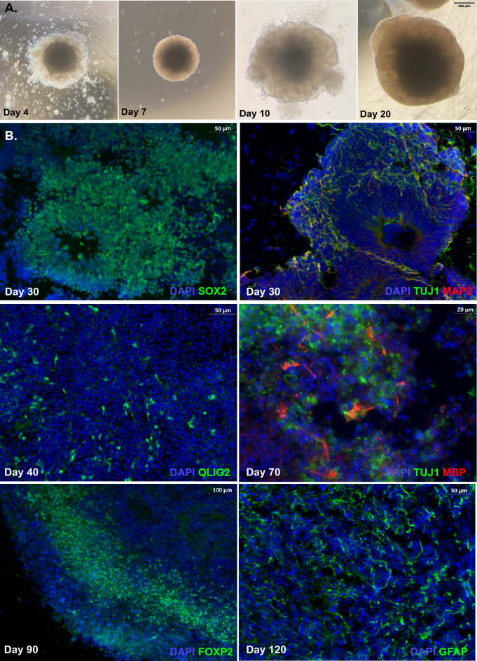
Figure 1.
Representative images from a Control human cerebral organoid (hCO). (A) Morphological changes of hCO under light microscope. By day 4 after differentiation, an embryoid body (EBs) with round and smooth edges form in the EB formation medium. By day 7, the EB develops brighter borders in the induction medium. The organoid embedded in Matrigel droplet shows budding of the surface around day 10. Organoids kept in the maturation medium exhibit dense core and optically translucent edges by day 20 and beyond. (B) Immunostaining of human cerebral organoids during development. Prototypical SOX2 + ventricular zone (VZ) at day 30, while TUJ1 + MAP2 + neurons migrate to the outer layer of VZ. By day 40, OLIG2 + OPCs expand after treatment of PDGF-AA and IGF1 for 12 days. OPCs are induced to MBP producing oligodendrocytes by T3 treatment. FOXP2 + cells form a layer along the organoid. GFAP + astrocytes are well-developed after long-term (120 days) maturation.

