Fig. 1.
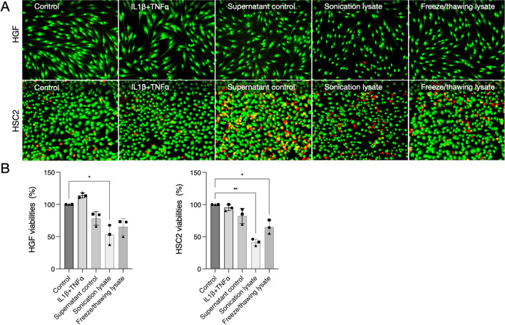
Cell viability was reduced after the exposure of cells to lysates. A The Live-dead assay shows the green-stained cells, which are viable, and the red-stained cells, which are dead. B The Live-dead results were confirmed by MTT colourimetric test. Sonication lysate provoked a cell viability reduction of about 50% in HGF and HSC2, which was a significant reduction compared to control or non-stimulated cells. The exposure of cells to IL1β and TNFα did not reduce cell viability in gingival fibroblasts and epithelial cells. Different symbol shapes mean independent experiments. * means p < 0.05 and ** means p < 0.01

