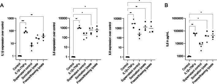
Fig. 2.
Interleukin expressions of HGF after exposure to IL1β + TNFα treatment or HSC2 lysates. A For gene expressions, HGF treated with supernatant control showed significantly less IL1β, IL6, and IL8 expressions than the treatment with the positive control for inflammation (IL1β + TNFα). On the other hand, HGF treated with sonication and freeze/thawing lysates expressed IL1β, IL6, and IL8 to similar levels of the IL1β + TNFα treatment. B IL6 immunoassay confirmed the elevated presence of interleukin produced by all groups in this in vitro model for inflammation. In the graphs, different symbol shapes mean independent experiments. * means p < 0.05 and ** means p < 0.01

