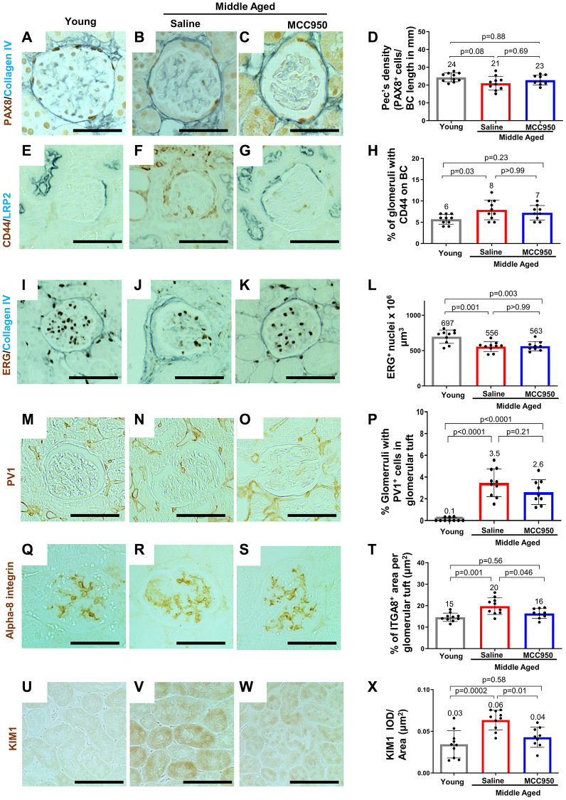
Figure 8.
Impact of MCC950 treatment on other glomerular cells and proximal tubules. Immunostainings comparing young to middle-aged saline- and MCC950-treated mice. (A–D) PAX8 (brown, nuclear) and Collagen IV staining (blue) was used to visualize parietal epithelial cells (PECs) and Bowman’s capsule, respectively. Data were quantified and PEC density was calculated by the ratio of PAX8-positive cells and Bowman’s capsule (BC) length (D). (E–H) Immunostaining for CD44 (brown) and LRP2 (blue) was used to label activated PECs and proximal tubules, respectively (E–G). To determine the number of glomeruli with activated PECs, data were quantified based on the presence of CD44 staining in BC (H). (I–L) Endothelial cell number was determined staining with the endothelial cell marker ERG (nuclear brown staining) and the BC marker Collagen IV (blue) (I–K). Data were quantified as number of nuclei per mm2 (L). (M–P) To assess endothelial injury, kidney sections were stained for PV-1 (brown) as a marker of injured endothelial cells (brown) and quantified as percentage of glomeruli with PV-1-positive cells (P). (Q–T) Alpha-8 integrin (ITGA8) staining (brown) was used as a measure of the mesangial area (brown) (Q–S) and quantified as the percent ITGA8-positive area within the glomerular tuft (T). (U–X) To determine the effect of MCC950 on proximal tubules, sections were stained with KIM1 (brown) (U–W) and quantified as IOD per area (X). In all panels representative images are shown and scale bars in the images correspond to 25 μm. In all the graphs, error bars are standard deviation, and the mean levels are stated by the number above the bars.

