FIGURE 5.

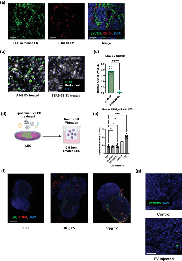
Lymphatic endothelial cells (LECs) are the LN recipient cells of EVs LECs regulate neutrophil recruitment and NETs formation. (a) Representative images of draining LNs after footpad B16F10 EV injection, showing LECs uptaking EVs Scale bars represent 20μm. (b) Representative images of LECs after A549 and BEAS‐2B EV treatment. Scale bars represent 100μm. (c) Quantification of the EV uptake (relative fluorescence area of CFSE to DAPI) in LECs. (d) Schematic illustration of the Boyden chamber transwell assay. (e) Quantification of Boyden chamber transwell assay, showing neutrophil migration towards CM from treated LECs. Data shown as mean ± SEM. *, P < 0.05; ***, P < 0.005; **** P < 0.001 by Mann–Whitney t test or One‐Way ANOVA. (f) Representative images of draining LNs after footpad PBS or different doses of B16F10 EV injection, indicating the subsequent neutrophil recruitment and NETs deposition. Scale bars represent 500 μm. (g) Representative images of draining LNs after footpad PBS or 10 μg B16F10 EV injection, indicating the increased expression of premetastatic marker VEGFR3. Scale bars represent 50 μm.
