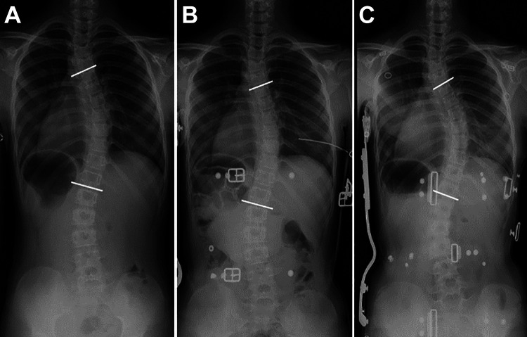
Figure 2.
A representative radiograph of a patient with in-brace deterioration (main curve: T5-12) showing the deterioration during brace treatment: (A) Pre-brace standing radiograph Cobb angle of 35°; (B) Initial inbrace radiograph Cobb angle of 29°; and (C) In-brace radiograph showing deterioration to Cobb angle of 44°.

