FIG. 1.
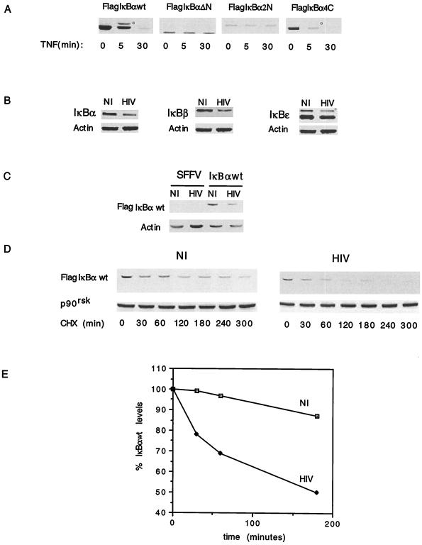
Functional characterization of Flag-IκBα molecules in U937 cells. (A) Pooled clones of U937 cells expressing the different Flag-IκBα constructs were stimulated with TNF for different time periods, and the cell lysates were analyzed by immunoblotting with anti-Flag antibodies. The hyperphosphorylated form of IκBα is indicated by a small circle. (B) Immunoblotting of cell lysates from mock-infected (NI) or HIV-infected (HIV), SFFV-expressing U937 cells with anti-IκBα, anti-IκBβ, anti-IκBɛ, and antiactin antibodies. The hyperphosphorylated form of IκBɛ is indicated by a small circle. (C) Immunoblotting of cell lysates from mock-infected (NI) or HIV-infected (HIV), SFFV- or Flag-IκBα-wt-expressing U937 cells with anti-Flag and antiactin antibodies. (D) The half-life of Flag-IκBα-wt was estimated by immunoblotting of cell lysates from mock-infected (NI) or HIV-infected (HIV), Flag-IκBα-wt-expressing U937 cells treated with cycloheximide (CHX) for different periods of time with anti-Flag antibodies. Equal protein loading was calculated by immunoblotting the same membrane with anti-p90rsk antibody. (E) The half-life of Flag-IκBα-wt was calculated by measuring with a densitometer the disintegrations per minute of Flag-IκBα-wt and normalizing them to those for p90rsk from each experimental time point shown in panel D.

