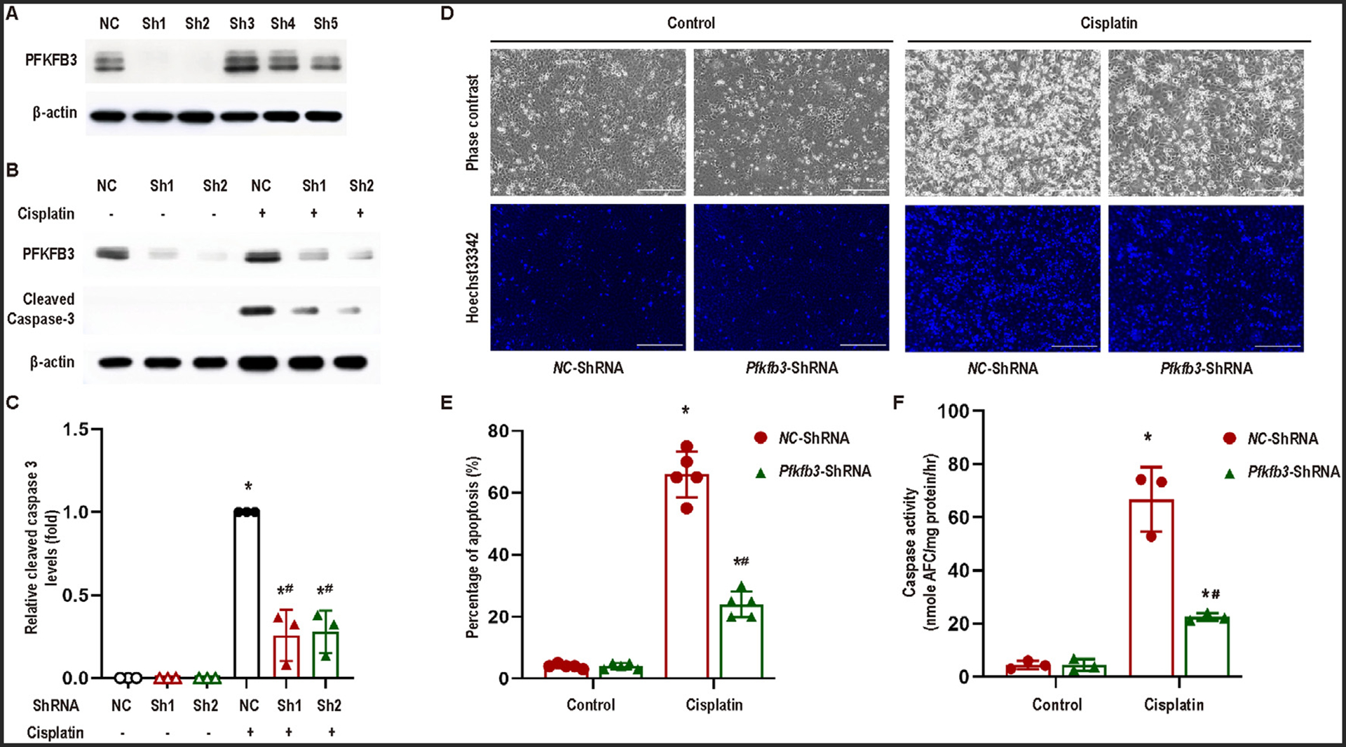
Fig 3.

Knockdown of PFKFB3 alleviates cisplatin-induced apoptosis in RPTCs. (A) RPTCs were transfected with different Pfkfb3-ShRNAs (Sh1–5) or negative control shRNA (NC), and then selected with puromycin for 2 weeks. Whole cell lysates were collected for immunoblot analysis of PFKFB3 and β-actin. The cells stably transfected with Pfkfb3-ShRNA1 were used for subsequent experiments. (B)−(F) Pfkfb3-ShRNA and negative control (NC) RPTCs were incubated with or without 20 μM cisplatin for 24 hours. (B) Immunoblot analysis for PFKFB3 and cleaved caspase-3 in Pfkfb3-knockdown and negative control RPTCs. β-Actin was used as the internal loading control. (C) Densitometry analysis of cleaved caspase-3 normalized by β-Actin signals (n = 3). (D) Representative cell and nuclear images after Hoechst 33342 staining. Scale bar = 200 μm. (E) Percentage of apoptosis assessed morphologically (n = 5). (F) Caspase activity. Data are expressed as means ± SD, n = 3. *, P < 0.05 vs control groups; #, P < 0.05 vs. cisplatin-treated NC-ShRNA RPTCs.
