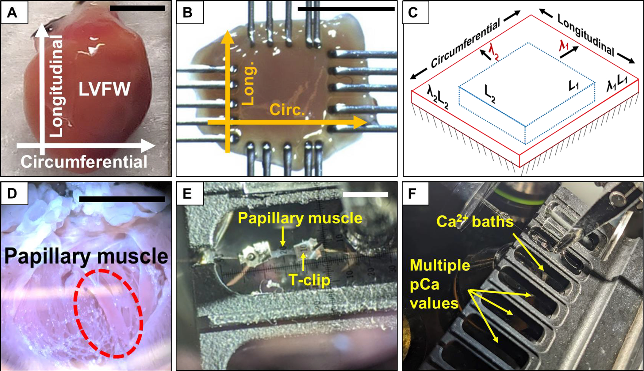
Fig. 2.

Overview of steps in the testing process. (A) Heart with designated orientations. (B) Mounted LVFW specimen post-skinning with orientations labeled. (C) Schematic of stretching and activation test protocol. Specimens were passively stretched to λ1 and λ2 in the circumferential and longitudinal directions, respectively, prior to active isometric contraction. (D) Image of an open heart for papillary muscle extraction. (E) Papillary muscle mounted to the testing device using aluminum T-clips prior to active isometric contraction. (F) Movement of mounted papillary muscle specimen through baths with different Ca2+ concentration levels. Scale bars in A, B, D, E: 5 mm.
