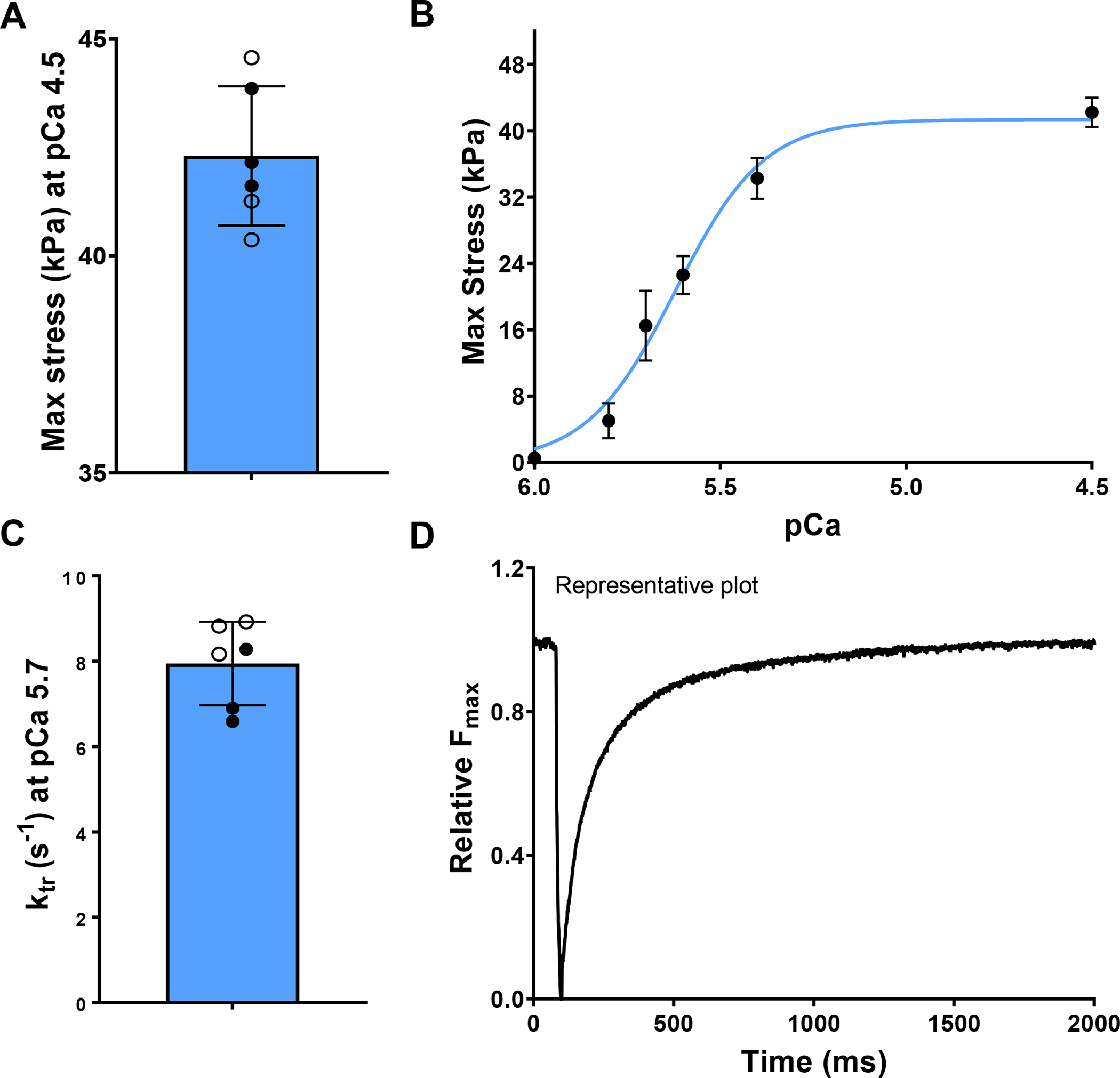
Fig. 8.

(A) The maximum contractile stress developed in the papillary muscle. (B) The effect of extrinsic Ca2+ concentration on maximum contractile stress (n = 6). (C) The rate of force development after cross-bridge dissociation, and (D) representative time for force development after cross-bridge dissociation. The filled and hollow markers indicate male and female specimens, respectively.
