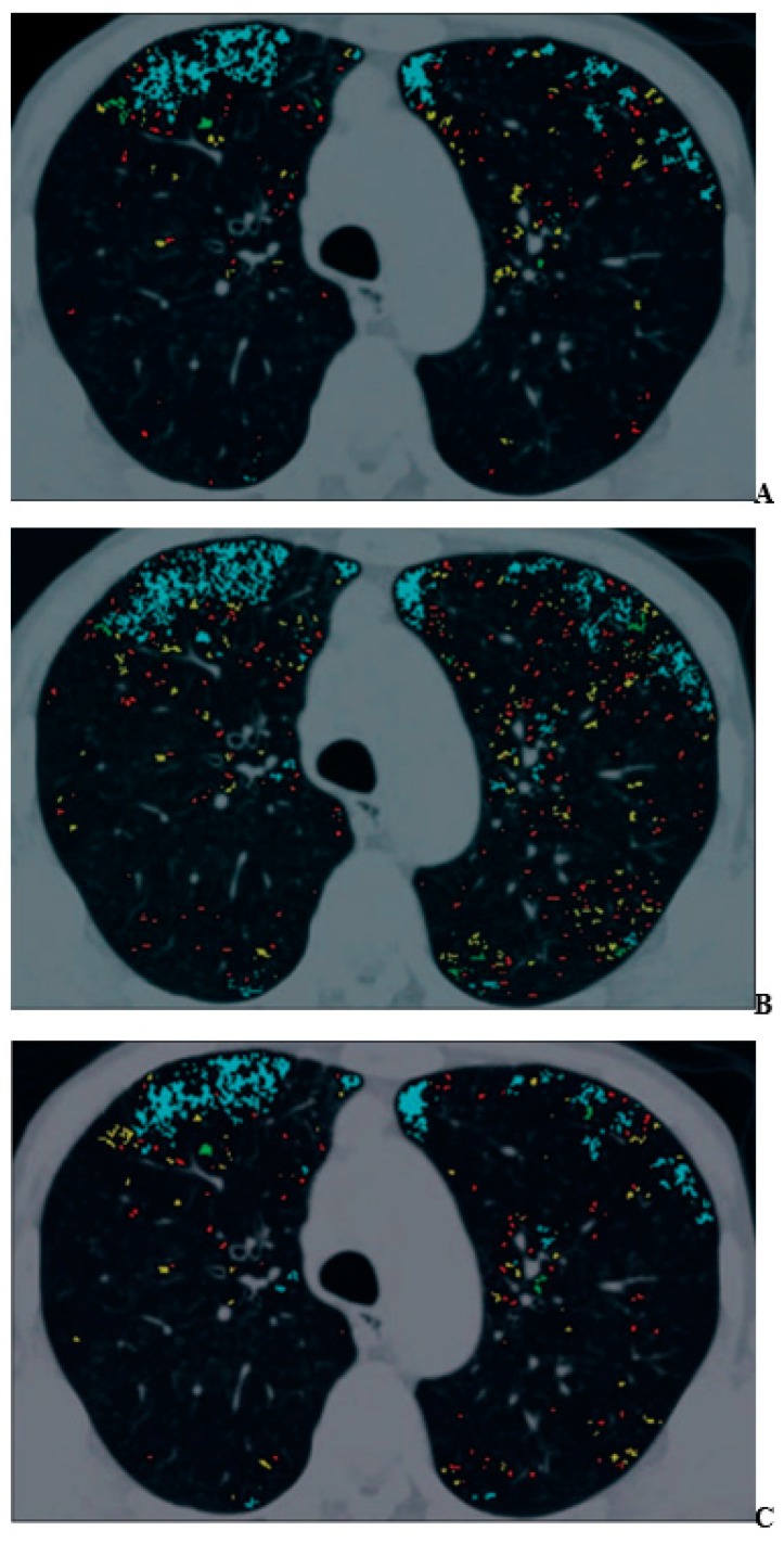
Figure 1.
A 70-year-old man with mild pulmonary emphysema (permission from reference [44]). (A–C) Images show low-attenuation lung regions with standard-dose CT (A), low-dose CT without adaptive iterative dose reduction using 3D processing (B), and low-dose CT with adaptive iterative dose reduction using 3D processing (C). Color coding of low-attenuation lung regions is as follows: class 1, red; class 2, yellow; class 3, green; and class 4, cyan.

