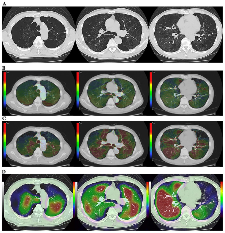
Figure 4.
A 75-year-old male smoker with “Moderate COPD” (58 pack-years, FEV1/FVC%: 55%, %FEV1: 58%) (permission from reference [108]). (A) (L to R: cranial to caudal): Thin-section CT shows a heterogeneously distributed low attenuation area due to pulmonary emphysema. (B) (L to R: cranial to caudal): DECT shows heterogeneous xenon enhancement within the lung and areas of reduced xenon enhancements corresponding well to the distribution of the areas of low attenuation. Total ventilation defect score was 66, and functional lung volume was 65.8%. (C) (L to R: cranial to caudal): Subtraction CT shows heterogeneous xenon enhancement within the lung and areas of reduced xenon enhancement corresponding well to the distribution of the areas of low attenuation. Total ventilation defect score was 88, and functional lung volume was 53.7%. (D) (L to R: cranial to caudal): Co-registered Kr–81m ventilation SPECT/CT shows markedly heterogeneous uptakes within the lung. Regional uptakes correspond well to areas of low attenuation. Total ventilation defect score was 84, and functional lung volume was 55.8%. Regional uptakes of Kr–81m show better correspondence to xenon enhancement on subtraction CT than on DECT.

