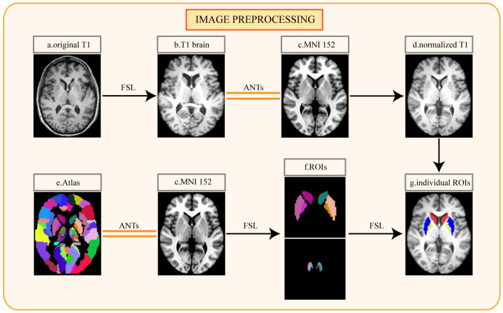
Figure 2.
Pipeline for image preprocessing: (a) original T1-weighted MRI of individual patient; (b) T1 image after cranium removal; (c) Montreal Neurosciences Institute 152 (MNI152) standard space; (d) normalized T1 image; (e) automated anatomical labeling atlas 3; (f) the regions of interest segmented from the atlas; (g) the regions of interest segmented from the individual normalized image. FSL, the FMRIB Software Library; ANTs, Advanced Normalization Tools.

