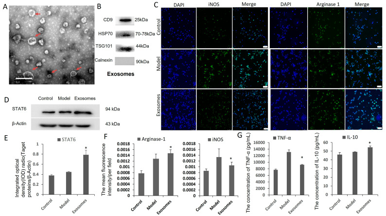
Figure 7.
ADSC-derived exosomes and macrophage polarization assay in vitro. (A) TEM analysis presented that ADSC-derived exosomes were round membrane-bound vesicles and the diameter is about 30 mm to 150 nm as indicated by the red arrow. Scale bar is 200 nm. (B) Exosomes from rabbit ADSCs were identified by markers CD9, HSP70, TSG101 and Calnexin by Western blot. (C,F) Immunofluorescence detection of M1, M2 macrophage marker proteins iNOS and arginase-1. Compared with the model group, iNOS level was lower, and arginase-1 expression was higher in ADSC exosome treatment group. Original magnification is 200. (D,E) Western blot detection showed the expression of STAT6 signal molecules increased significantly in ADSC-derived exosome group. (G) ELISA results suggested that concentration of cytokine TNF-α decreased and IL-10 increased in ADSC exosomes group. * p < 0.05.

