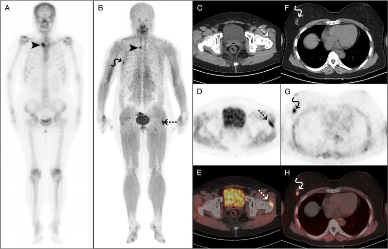
FIGURE 1.

68Ga-FAPI-04 PET/CT detected left hip synovitis in a 57-year-old woman with SAPHO syndrome. The woman presented with anterior chest wall and left hip pain for 6 months and palmar pustules over the past 3 years. Laboratory examination showed elevated ESR (67 mm/h) and hs-CRP (13 mg/L) and a positive ANA test. 99mTc-MDP bone scan (A) and 68Ga-FAPI-04 PET/CT (B) both showed increased tracer uptake in the right sternoclavicular joint (arrowheads). PET/CT (B–E) also showed increased tracer uptake in the synovial sac of the left hip joint (dotted arrows). Subsequent ultrasonography of the left hip confirmed the presence of synovitis. Furthermore, a nodule in the right breast with abnormal tracer uptake (F–H, curved arrows) was subsequently confirmed as lobular carcinoma in situ via puncture.
